A structural mechanism for directing corepressor-selective inverse agonism of PPAR gamma.
Brust, R., Shang, J., Fuhrmann, J., Mosure, S.A., Bass, J., Cano, A., Heidari, Z., Chrisman, I.M., Nemetchek, M.D., Blayo, A.L., Griffin, P.R., Kamenecka, T.M., Hughes, T.S., Kojetin, D.J.(2018) Nat Commun 9: 4687-4687
- PubMed: 30409975 Search on PubMedSearch on PubMed Central
- DOI: https://doi.org/10.1038/s41467-018-07133-w
- Primary Citation Related Structures:
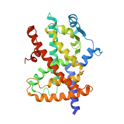
6C1I - PubMed Abstract:
Small chemical modifications can have significant effects on ligand efficacy and receptor activity, but the underlying structural mechanisms can be difficult to predict from static crystal structures alone. Here we show how a simple phenyl-to-pyridyl substitution between two common covalent orthosteric ligands targeting peroxisome proliferator-activated receptor (PPAR) gamma converts a transcriptionally neutral antagonist (GW9662) into a repressive inverse agonist (T0070907) relative to basal cellular activity. X-ray crystallography, molecular dynamics simulations, and mutagenesis coupled to activity assays reveal a water-mediated hydrogen bond network linking the T0070907 pyridyl group to Arg288 that is essential for corepressor-selective inverse agonism. NMR spectroscopy reveals that PPARγ exchanges between two long-lived conformations when bound to T0070907 but not GW9662, including a conformation that prepopulates a corepressor-bound state, priming PPARγ for high affinity corepressor binding. Our findings demonstrate that ligand engagement of Arg288 may provide routes for developing corepressor-selective repressive PPARγ ligands.
- Department of Integrative Structural and Computational Biology, The Scripps Research Institute, Jupiter, FL, 33458, USA.
Organizational Affiliation: