6C08

Zebrafish SLC38A9 with arginine bound in the cytosol open state


Experimental Data Snapshot

  • Method: X-RAY DIFFRACTION
  • Resolution: 3.17 Å
  • R-Value Free: 0.285 
  • R-Value Work: 0.267 
  • R-Value Observed: 0.267 

wwPDB Validation   3D Report Full Report


This is version 1.0 of the entry. See complete history


Literature

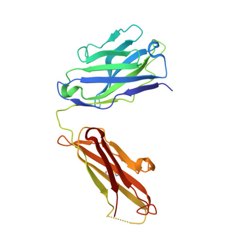
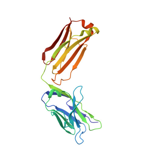
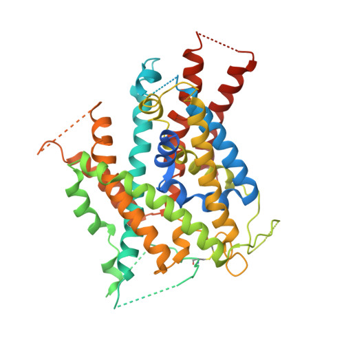
Crystal structure of arginine-bound lysosomal transporter SLC38A9 in the cytosol-open state.

Lei, H.T.Ma, J.Sanchez Martinez, S.Gonen, T.

(2018) Nat Struct Mol Biol 25: 522-527

  • DOI: https://doi.org/10.1038/s41594-018-0072-2
  • Primary Citation of Related Structures:  
    6C08

  • PubMed Abstract: 

    Recent advances in understanding intracellular amino acid transport and mechanistic target of rapamycin complex 1 (mTORC1) signaling shed light on solute carrier 38, family A member 9 (SLC38A9), a lysosomal transporter responsible for the binding and translocation of several essential amino acids. Here we present the first crystal structure of SLC38A9 from Danio rerio in complex with arginine. As captured in the cytosol-open state, the bound arginine was locked in a transitional state stabilized by transmembrane helix 1 (TM1) of drSLC38A9, which was anchored at the groove between TM5 and TM7. These anchoring interactions were mediated by the highly conserved WNTMM motif in TM1, and mutations in this motif abolished arginine transport by drSLC38A9. The underlying mechanism of substrate binding is critical for sensitizing the mTORC1 signaling pathway to amino acids and for maintenance of lysosomal amino acid homeostasis. This study offers a first glimpse into a prototypical model for SLC38 transporters.


  • Organizational Affiliation

    Janelia Research Campus, Howard Hughes Medical Institute, Ashburn, VA, USA.


Macromolecules
Find similar proteins by:  (by identity cutoff)  |  3D Structure
Entity ID: 1
MoleculeChains Sequence LengthOrganismDetailsImage
antibody Fab Heavy Chain
A, D
218Mus musculusMutation(s): 0 
Entity Groups  
Sequence Clusters30% Identity50% Identity70% Identity90% Identity95% Identity100% Identity
Sequence Annotations
Expand
  • Reference Sequence
Find similar proteins by:  (by identity cutoff)  |  3D Structure
Entity ID: 2
MoleculeChains Sequence LengthOrganismDetailsImage
antibody Fab light chain
B, E
215Mus musculusMutation(s): 0 
Entity Groups  
Sequence Clusters30% Identity50% Identity70% Identity90% Identity95% Identity100% Identity
Sequence Annotations
Expand
  • Reference Sequence
Find similar proteins by:  (by identity cutoff)  |  3D Structure
Entity ID: 3
MoleculeChains Sequence LengthOrganismDetailsImage
Sodium-coupled neutral amino acid transporter 9
C, F
479Danio rerioMutation(s): 4 
Gene Names: slc38a9zgc:154088
Membrane Entity: Yes 
UniProt
Find proteins for Q08BA4 (Danio rerio)
Explore Q08BA4 
Go to UniProtKB:  Q08BA4
Entity Groups  
Sequence Clusters30% Identity50% Identity70% Identity90% Identity95% Identity100% Identity
UniProt GroupQ08BA4
Sequence Annotations
Expand
  • Reference Sequence
Small Molecules
Ligands 1 Unique
IDChains Name / Formula / InChI Key2D Diagram3D Interactions
ARG
Query on ARG

Download Ideal Coordinates CCD File 
G [auth F]ARGININE
C6 H15 N4 O2
ODKSFYDXXFIFQN-BYPYZUCNSA-O
Experimental Data & Validation

Experimental Data

  • Method: X-RAY DIFFRACTION
  • Resolution: 3.17 Å
  • R-Value Free: 0.285 
  • R-Value Work: 0.267 
  • R-Value Observed: 0.267 
  • Space Group: P 1 21 1
Unit Cell:
Length ( Å )Angle ( ˚ )
a = 136.613α = 90
b = 82.808β = 100.02
c = 158.922γ = 90
Software Package:
Software NamePurpose
PHENIXrefinement
Aimlessdata scaling
PDB_EXTRACTdata extraction
XDSdata reduction
PHASERphasing

Structure Validation

View Full Validation Report


Entry History 

Deposition Data

Revision History  (Full details and data files)

  • Version 1.0: 2018-06-20
    Type: Initial release