6C02

Human ectonucleotide pyrophosphatase / phosphodiesterase 3 (ENPP3, NPP3, CD203c), inactive (T205A), N594S, with alpha,beta-methylene-ATP (AMPCPP)


Experimental Data Snapshot

  • Method: X-RAY DIFFRACTION
  • Resolution: 1.94 Å
  • R-Value Free: 0.197 
  • R-Value Work: 0.166 
  • R-Value Observed: 0.168 

wwPDB Validation   3D Report Full Report


Ligand Structure Quality Assessment 


This is version 2.1 of the entry. See complete history


Literature

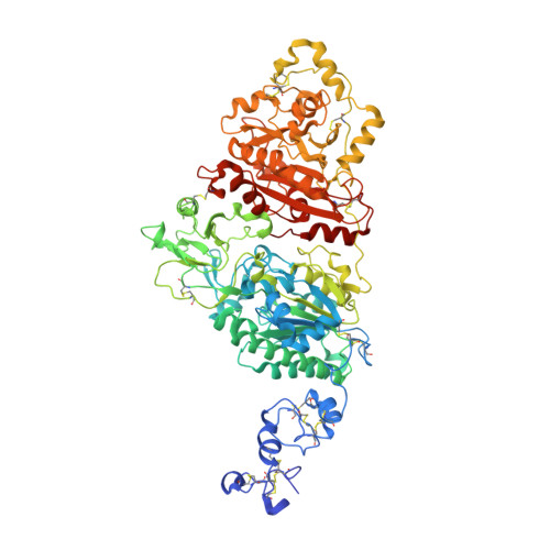
Structural basis for nucleotide recognition by the ectoenzyme CD203c.

Gorelik, A.Randriamihaja, A.Illes, K.Nagar, B.

(2018) FEBS J 285: 2481-2494

  • DOI: https://doi.org/10.1111/febs.14489
  • Primary Citation of Related Structures:  
    6C01, 6C02

  • PubMed Abstract: 

    The ecto-nucleotide pyrophosphatase/phosphodiesterase (NPP) enzyme family modulates purinergic signaling by degrading extracellular nucleotides. CD203c (NPP3, ENPP3) regulates the inflammatory response of basophils via ATP hydrolysis and is a marker for allergen sensitivity on the surface of these cells. Multiple other roles and substrates have also been proposed for this protein. In order to gain insight into its molecular functions, we determined the crystal structure of human NPP3 as well as its complex with an ATP analog. The enzyme exhibits little preference for nucleobase type, and forms specific contacts with the alpha and beta phosphate groups of its ligands. Dimerization of the protein does not affect its catalytic activity. These findings expand our understanding of substrate recognition within the NPP family.


  • Organizational Affiliation

    Department of Biochemistry and Groupe de Recherche Axé sur la Structure des Protéines, McGill University, Montreal, Quebec, Canada.


Macromolecules
Find similar proteins by:  (by identity cutoff)  |  3D Structure
Entity ID: 1
MoleculeChains Sequence LengthOrganismDetailsImage
Ectonucleotide pyrophosphatase/phosphodiesterase family member 3
A, B
838Homo sapiensMutation(s): 2 
Gene Names: ENPP3PDNP3
EC: 3.1.4.1 (PDB Primary Data), 3.6.1.9 (PDB Primary Data)
UniProt & NIH Common Fund Data Resources
Find proteins for O14638 (Homo sapiens)
Explore O14638 
Go to UniProtKB:  O14638
PHAROS:  O14638
GTEx:  ENSG00000154269 
Entity Groups  
Sequence Clusters30% Identity50% Identity70% Identity90% Identity95% Identity100% Identity
UniProt GroupO14638
Sequence Annotations
Expand
  • Reference Sequence
Oligosaccharides

Help

Entity ID: 2
MoleculeChains Length2D Diagram Glycosylation3D Interactions
2-acetamido-2-deoxy-beta-D-glucopyranose-(1-4)-2-acetamido-2-deoxy-beta-D-glucopyranose
C, F, J, L, M
2N-Glycosylation
Glycosylation Resources
GlyTouCan:  G42666HT
GlyCosmos:  G42666HT
GlyGen:  G42666HT
Entity ID: 3
MoleculeChains Length2D Diagram Glycosylation3D Interactions
beta-D-mannopyranose-(1-4)-2-acetamido-2-deoxy-beta-D-glucopyranose-(1-4)-2-acetamido-2-deoxy-beta-D-glucopyranose
D, K
3N-Glycosylation
Glycosylation Resources
GlyTouCan:  G15407YE
GlyCosmos:  G15407YE
GlyGen:  G15407YE
Entity ID: 4
MoleculeChains Length2D Diagram Glycosylation3D Interactions
2-acetamido-2-deoxy-beta-D-glucopyranose-(1-4)-[alpha-L-fucopyranose-(1-6)]2-acetamido-2-deoxy-beta-D-glucopyranose
E, G, N
3N-Glycosylation
Glycosylation Resources
GlyTouCan:  G21290RB
GlyCosmos:  G21290RB
GlyGen:  G21290RB
Entity ID: 5
MoleculeChains Length2D Diagram Glycosylation3D Interactions
alpha-L-fucopyranose-(1-6)-2-acetamido-2-deoxy-beta-D-glucopyranose
H, I
2N-Glycosylation
Glycosylation Resources
GlyTouCan:  G86851RC
GlyCosmos:  G86851RC
GlyGen:  G86851RC
Small Molecules
Ligands 6 Unique
IDChains Name / Formula / InChI Key2D Diagram3D Interactions
APC
Query on APC

Download Ideal Coordinates CCD File 
EA [auth B],
X [auth A]
DIPHOSPHOMETHYLPHOSPHONIC ACID ADENOSYL ESTER
C11 H18 N5 O12 P3
CAWZRIXWFRFUQB-IOSLPCCCSA-N
SO4
Query on SO4

Download Ideal Coordinates CCD File 
CA [auth B],
DA [auth B],
V [auth A],
W [auth A]
SULFATE ION
O4 S
QAOWNCQODCNURD-UHFFFAOYSA-L
ZN
Query on ZN

Download Ideal Coordinates CCD File 
O [auth A],
P [auth A],
Y [auth B],
Z [auth B]
ZINC ION
Zn
PTFCDOFLOPIGGS-UHFFFAOYSA-N
CA
Query on CA

Download Ideal Coordinates CCD File 
AA [auth B],
Q [auth A]
CALCIUM ION
Ca
BHPQYMZQTOCNFJ-UHFFFAOYSA-N
CL
Query on CL

Download Ideal Coordinates CCD File 
S [auth A],
T [auth A],
U [auth A]
CHLORIDE ION
Cl
VEXZGXHMUGYJMC-UHFFFAOYSA-M
NA
Query on NA

Download Ideal Coordinates CCD File 
BA [auth B],
R [auth A]
SODIUM ION
Na
FKNQFGJONOIPTF-UHFFFAOYSA-N
Experimental Data & Validation

Experimental Data

  • Method: X-RAY DIFFRACTION
  • Resolution: 1.94 Å
  • R-Value Free: 0.197 
  • R-Value Work: 0.166 
  • R-Value Observed: 0.168 
  • Space Group: P 1 21 1
Unit Cell:
Length ( Å )Angle ( ˚ )
a = 78.656α = 90
b = 169.457β = 103.87
c = 79.062γ = 90
Software Package:
Software NamePurpose
PHENIXrefinement
HKL-2000data reduction
HKL-2000data scaling
PHASERphasing

Structure Validation

View Full Validation Report


Ligand Structure Quality Assessment 


Entry History & Funding Information

Deposition Data


Funding OrganizationLocationGrant Number
Canadian Institutes of Health Research (CIHR)CanadaMOP-133535

Revision History  (Full details and data files)

  • Version 1.0: 2018-05-02
    Type: Initial release
  • Version 1.1: 2018-06-13
    Changes: Data collection, Database references
  • Version 1.2: 2018-07-18
    Changes: Data collection, Database references
  • Version 1.3: 2020-01-08
    Changes: Author supporting evidence, Data collection
  • Version 2.0: 2020-07-29
    Type: Remediation
    Reason: Carbohydrate remediation
    Changes: Atomic model, Data collection, Derived calculations, Structure summary
  • Version 2.1: 2023-10-04
    Changes: Data collection, Database references, Derived calculations, Refinement description, Structure summary