


Crystal Structure of Glucose-6-phosphate Isomerase from Elizabethkingia anophelis with bound Glucose-6-phosphate
Dranow, D.M., Mayclin, S.J., Lorimer, D.D., Edwards, T.E.To be published.
Experimental Data Snapshot
Starting Model: experimental
View more details
Entity ID: 1 | |||||
|---|---|---|---|---|---|
| Molecule | Chains | Sequence Length | Organism | Details | Image |
| Glucose-6-phosphate isomerase | 555 | Elizabethkingia anophelis NUHP1 | Mutation(s): 0 Gene Names: pgi, BD94_3890 EC: 5.3.1.9 | 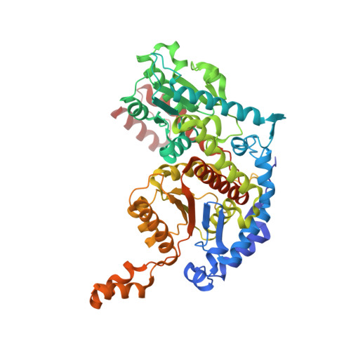 | |
UniProt | |||||
Find proteins for A0A077EQ93 (Elizabethkingia anophelis NUHP1) Explore A0A077EQ93 Go to UniProtKB: A0A077EQ93 | |||||
Entity Groups | |||||
| Sequence Clusters | 30% Identity50% Identity70% Identity90% Identity95% Identity100% Identity | ||||
| UniProt Group | A0A077EQ93 | ||||
Sequence AnnotationsExpand | |||||
Reference Sequence | |||||
| Ligands 3 Unique | |||||
|---|---|---|---|---|---|
| ID | Chains | Name / Formula / InChI Key | 2D Diagram | 3D Interactions | |
| G6P Download:Ideal Coordinates CCD File | C [auth A], H [auth B] | 6-O-phosphono-alpha-D-glucopyranose C6 H13 O9 P NBSCHQHZLSJFNQ-DVKNGEFBSA-N |  | ||
| GOL Download:Ideal Coordinates CCD File | G [auth A] | GLYCEROL C3 H8 O3 PEDCQBHIVMGVHV-UHFFFAOYSA-N |  | ||
| EDO Download:Ideal Coordinates CCD File | D [auth A], E [auth A], F [auth A], I [auth B] | 1,2-ETHANEDIOL C2 H6 O2 LYCAIKOWRPUZTN-UHFFFAOYSA-N |  | ||
| Length ( Å ) | Angle ( ˚ ) |
|---|---|
| a = 69.28 | α = 90 |
| b = 102.14 | β = 90 |
| c = 153.73 | γ = 90 |
| Software Name | Purpose |
|---|---|
| XDS | data reduction |
| XSCALE | data scaling |
| PHASER | phasing |
| PHENIX | refinement |
| PDB_EXTRACT | data extraction |


