6BX4

The crystal structure of fluoride channel Fluc Ec2 with Monobody S9


Experimental Data Snapshot

  • Method: X-RAY DIFFRACTION
  • Resolution: 2.55 Å
  • R-Value Free: 0.246 
  • R-Value Work: 0.222 
  • R-Value Observed: 0.223 

wwPDB Validation   3D Report Full Report


Ligand Structure Quality Assessment 


This is version 1.3 of the entry. See complete history


Literature

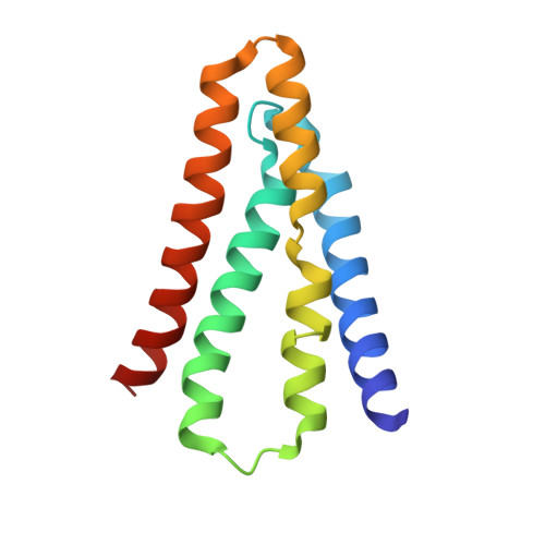
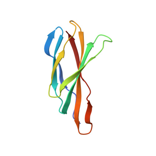
Molecular Interactions between a Fluoride Ion Channel and Synthetic Protein Blockers.

Turman, D.L.Cheloff, A.Z.Corrado, A.D.Nathanson, J.T.Miller, C.

(2018) Biochemistry 57: 1212-1218

  • DOI: https://doi.org/10.1021/acs.biochem.7b01272
  • Primary Citation of Related Structures:  
    6BX4, 6BX5

  • PubMed Abstract: 

    Fluoride ion channels of the Fluc family selectively export F - ions to rescue unicellular organisms from acute F - toxicity. Crystal structures of bacterial Fluc channels in complex with synthetic monobodies, fibronectin-derived soluble β-sandwich fold proteins, show 2-fold symmetric homodimers with an antiparallel transmembrane topology. Monobodies also block Fluc F - current via a pore blocking mechanism. However, little is known about the energetic contributions of individual monobody residues to the affinity of the monobody-channel complex or whether the structural paratope corresponds to functional reality. This study seeks to structurally identify and compare residues interacting with Fluc between two highly similar monobodies and subjects them to mutagenesis and functional measurements of equilibrium affinities via a fluorescence anisotropy binding assay to determine their energetic contributions. The results indicate that the functional and structural paratopes strongly agree and that many Tyr residues at the interface, while playing a key role in affinity, can be substituted with Phe and Trp without large disruptions.


  • Organizational Affiliation

    Department of Biochemistry, Howard Hughes Medical Institute, Brandeis University , 415 South Street, Waltham, Massachusetts 02453, United States.


Macromolecules
Find similar proteins by:  (by identity cutoff)  |  3D Structure
Entity ID: 1
MoleculeChains Sequence LengthOrganismDetailsImage
Fluoride ion transporter CrcB
A, B
123Escherichia coliMutation(s): 1 
Gene Names: 
Membrane Entity: Yes 
UniProt
Find proteins for Q6J5N4 (Escherichia coli)
Explore Q6J5N4 
Go to UniProtKB:  Q6J5N4
Entity Groups  
Sequence Clusters30% Identity50% Identity70% Identity90% Identity95% Identity100% Identity
UniProt GroupQ6J5N4
Sequence Annotations
Expand
  • Reference Sequence
Find similar proteins by:  (by identity cutoff)  |  3D Structure
Entity ID: 2
MoleculeChains Sequence LengthOrganismDetailsImage
Monobody
C, D
96Homo sapiensMutation(s): 0 
Entity Groups  
Sequence Clusters30% Identity50% Identity70% Identity90% Identity95% Identity100% Identity
Sequence Annotations
Expand
  • Reference Sequence
Experimental Data & Validation

Experimental Data

  • Method: X-RAY DIFFRACTION
  • Resolution: 2.55 Å
  • R-Value Free: 0.246 
  • R-Value Work: 0.222 
  • R-Value Observed: 0.223 
  • Space Group: P 41
Unit Cell:
Length ( Å )Angle ( ˚ )
a = 87.412α = 90
b = 87.412β = 90
c = 144.333γ = 90
Software Package:
Software NamePurpose
REFMACrefinement
XDSdata reduction
Aimlessdata scaling
PHASERphasing
PDB_EXTRACTdata extraction

Structure Validation

View Full Validation Report


Ligand Structure Quality Assessment 


Entry History & Funding Information

Deposition Data


Funding OrganizationLocationGrant Number
National Institutes of Health/National Institute of General Medical Sciences (NIH/NIGMS)United StatesR01-GM10723

Revision History  (Full details and data files)

  • Version 1.0: 2018-02-14
    Type: Initial release
  • Version 1.1: 2018-02-28
    Changes: Database references
  • Version 1.2: 2020-01-01
    Changes: Author supporting evidence
  • Version 1.3: 2023-10-04
    Changes: Data collection, Database references, Derived calculations, Refinement description