6BVC | pdb_00006bvc

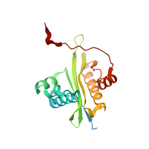
Crystal structure of AAC(3)-Ia in complex with coenzyme A


Experimental Data Snapshot

  • Method: X-RAY DIFFRACTION
  • Resolution: 1.81 Å
  • R-Value Free: 
    0.191 (Depositor), 0.189 (DCC) 
  • R-Value Work: 
    0.162 (Depositor), 0.163 (DCC) 
  • R-Value Observed: 
    0.164 (Depositor) 

Starting Model: experimental
View more details

wwPDB Validation 3D Report Full Report

Validation slider image for 6BVC

Ligand Structure Quality Assessment 


This is version 1.5 of the entry. See complete history

Literature

Structure of the full-length Serratia marcescens acetyltransferase AAC(3)-Ia in complex with coenzyme A.

Popov, G.Evdokimova, E.Stogios, P.J.Savchenko, A.

(2020) Protein Sci 29: 803-808

  • DOI: https://doi.org/10.1002/pro.3811
  • Primary Citation Related Structures: 
    6BVC

  • PubMed Abstract: 

    Acyl-coenzyme A-dependent N-acetyltransferases (AACs) catalyze the modification of aminoglycosides rendering the bacteria carrying such enzymes resistant to this class of antibiotics. Here we present the crystal structure of AAC(3)-Ia enzyme from Serratia marcescens in complex with coenzyme A determined to 1.8 Å resolution. This enzyme served as an architype for the AAC enzymes targeting the amino group at Position 3 of aminoglycoside main aminocyclitol ring. The structure of this enzyme has been previously determined only in truncated form and was interpreted as distinct from subsequently characterized AACs. The reason for the unusual arrangement of secondary structure elements of AAC(3)-Ia was not further investigated. By determining the full-length structure of AAC(3)-Ia we establish that this enzyme adopts the canonical AAC fold conserved across this family and it does not undergo through significant rearrangement of secondary structure elements upon ligand binding as was proposed previously. In addition, our results suggest that the C-terminal tail in AAC(3)-Ia monomer forms intramolecular hydrogen bonds that contributes to formation of stable dimer, representing the predominant oligomeric state for this enzyme.


  • Organizational Affiliation
    • Department of Microbiology, Immunology and Infectious Diseases, University of Calgary, Calgary, Alberta, Canada.

Macromolecule Content 

  • Total Structure Weight: 21.78 kDa 
  • Atom Count: 1,498 
  • Modeled Residue Count: 154 
  • Deposited Residue Count: 177 
  • Unique protein chains: 1

Macromolecules

Find similar proteins by:|  3D Structure
Entity ID: 1
MoleculeChains  Sequence LengthOrganismDetailsImage
Aminoglycoside-(3)-N-acetyltransferase177Serratia marcescensMutation(s): 0 
Gene Names: aacC1
UniProt
Find proteins for Q53396 (Serratia marcescens)
Explore Q53396 
Go to UniProtKB:  Q53396
Entity Groups
Sequence Clusters30% Identity50% Identity70% Identity90% Identity95% Identity100% Identity
UniProt GroupQ53396
Sequence Annotations
Expand
Reference Sequence

Small Molecules

Ligands 4 Unique
IDChains Name / Formula / InChI Key2D Diagram3D Interactions
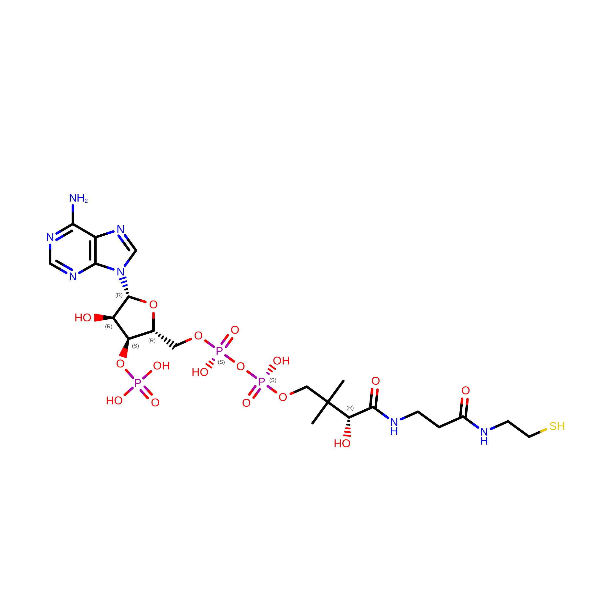
COA

Query on COA


Download:Ideal Coordinates CCD File
B [auth A]COENZYME A
C21 H36 N7 O16 P3 S
RGJOEKWQDUBAIZ-IBOSZNHHSA-N
PE3

Query on PE3


Download:Ideal Coordinates CCD File
D [auth A],
E [auth A]
3,6,9,12,15,18,21,24,27,30,33,36,39-TRIDECAOXAHENTETRACONTANE-1,41-DIOL
C28 H58 O15
ILLKMACMBHTSHP-UHFFFAOYSA-N
GOL

Query on GOL


Download:Ideal Coordinates CCD File
F [auth A],
G [auth A],
H [auth A]
GLYCEROL
C3 H8 O3
PEDCQBHIVMGVHV-UHFFFAOYSA-N
CL

Query on CL


Download:Ideal Coordinates CCD File
C [auth A]CHLORIDE ION
Cl
VEXZGXHMUGYJMC-UHFFFAOYSA-M

Experimental Data & Validation

Experimental Data

  • Method: X-RAY DIFFRACTION
  • Resolution: 1.81 Å
  • R-Value Free:  0.191 (Depositor), 0.189 (DCC) 
  • R-Value Work:  0.162 (Depositor), 0.163 (DCC) 
  • R-Value Observed: 0.164 (Depositor) 
Space Group: C 2 2 2
Unit Cell:
Length ( Å )Angle ( ˚ )
a = 79.028α = 90
b = 100.875β = 90
c = 53.031γ = 90
Software Package:
Software NamePurpose
PHENIXrefinement
XDSdata reduction
Aimlessdata scaling
PHENIXphasing
PHENIXmodel building
Cootmodel building

Structure Validation

View Full Validation Report


Ligand Structure Quality Assessment 


Entry History 

& Funding Information

Deposition Data


Funding OrganizationLocationGrant Number
National Institutes of Health/National Institute Of Allergy and Infectious Diseases (NIH/NIAID)United StatesHHSN272201200026C
National Institutes of Health/National Institute Of Allergy and Infectious Diseases (NIH/NIAID)United StatesHHSN272201700060C

Revision History  (Full details and data files)

  • Version 1.0: 2017-12-27
    Type: Initial release
  • Version 1.1: 2018-01-03
    Changes: Structure summary
  • Version 1.2: 2018-01-17
    Changes: Author supporting evidence
  • Version 1.3: 2019-12-18
    Changes: Author supporting evidence
  • Version 1.4: 2023-10-04
    Changes: Data collection, Database references, Refinement description
  • Version 1.5: 2026-08-19
    Changes: Database references, Structure summary