6BUX | pdb_00006bux

CRYSTAL STRUCTURE OF APOBEC3G CATALYTIC DOMAIN COMPLEX WITH SUBSTRATE SSDNA


Experimental Data Snapshot

  • Method: X-RAY DIFFRACTION
  • Resolution: 1.86 Å
  • R-Value Free: 
    0.211 (Depositor), 0.210 (DCC) 
  • R-Value Work: 
    0.179 (Depositor), 0.180 (DCC) 
  • R-Value Observed: 
    0.180 (Depositor) 

Starting Model: experimental
View more details

wwPDB Validation 3D Report Full Report

Validation slider image for 6BUX

This is version 1.2 of the entry. See complete history

Literature

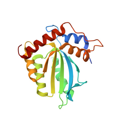
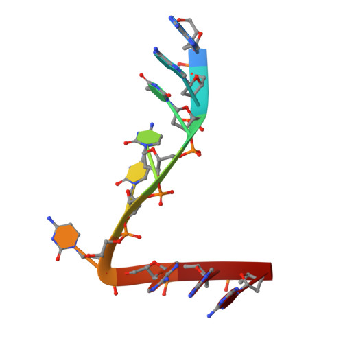
Crystal structure of the catalytic domain of HIV-1 restriction factor APOBEC3G in complex with ssDNA.

Maiti, A.Myint, W.Kanai, T.Delviks-Frankenberry, K.Sierra Rodriguez, C.Pathak, V.K.Schiffer, C.A.Matsuo, H.

(2018) Nat Commun 9: 2460-2460

  • DOI: https://doi.org/10.1038/s41467-018-04872-8
  • Primary Citation Related Structures: 
    6BUX

  • PubMed Abstract: 

    The human APOBEC3G protein is a cytidine deaminase that generates cytidine to deoxy-uridine mutations in single-stranded DNA (ssDNA), and capable of restricting replication of HIV-1 by generating mutations in viral genome. The mechanism by which APOBEC3G specifically deaminates 5'-CC motifs has remained elusive since structural studies have been hampered due to apparently weak ssDNA binding of the catalytic domain of APOBEC3G. We overcame the problem by generating a highly active variant with higher ssDNA affinity. Here, we present the crystal structure of this variant complexed with a ssDNA substrate at 1.86 Å resolution. This structure reveals atomic-level interactions by which APOBEC3G recognizes a functionally-relevant 5'-TCCCA sequence. This complex also reveals a key role of W211 in substrate recognition, implicating a similar recognition in activation-induced cytidine deaminase (AID) with a conserved tryptophan.


  • Organizational Affiliation
    • Basic Science Program, Leidos Biomedical Research, Inc., Frederick National Laboratory for Cancer Research, Frederick, MD, 21702, USA.

Macromolecule Content 

  • Total Structure Weight: 25.91 kDa 
  • Atom Count: 1,893 
  • Modeled Residue Count: 199 
  • Deposited Residue Count: 208 
  • Unique protein chains: 1
  • Unique nucleic acid chains: 1

Macromolecules


Find similar proteins by:|  3D Structure
Entity ID: 1
MoleculeChains  Sequence LengthOrganismDetailsImage
Apolipoprotein B mRNA editing enzyme catalytic subunit 3G catalytic domain199Homo sapiensMutation(s): 0 
EC: 3.5.4.38
UniProt & NIH Common Fund Data Resources
Find proteins for Q9HC16 (Homo sapiens)
Explore Q9HC16 
Go to UniProtKB:  Q9HC16
PHAROS:  Q9HC16
GTEx:  ENSG00000239713 
Entity Groups
Sequence Clusters30% Identity50% Identity70% Identity90% Identity95% Identity100% Identity
UniProt GroupQ9HC16
Sequence Annotations
Expand
Reference Sequence
Find similar nucleic acids by:  Sequence
Entity ID: 2
MoleculeChains LengthOrganismImage
DNA (5'-D(*AP*AP*TP*CP*CP*CP*AP*AP*A)-3')9synthetic construct
Sequence Annotations
Expand
Reference Sequence

Experimental Data & Validation

Experimental Data

  • Method: X-RAY DIFFRACTION
  • Resolution: 1.86 Å
  • R-Value Free:  0.211 (Depositor), 0.210 (DCC) 
  • R-Value Work:  0.179 (Depositor), 0.180 (DCC) 
  • R-Value Observed: 0.180 (Depositor) 
Space Group: P 1 21 1
Unit Cell:
Length ( Å )Angle ( ˚ )
a = 47.413α = 90
b = 47.237β = 103.13
c = 51.462γ = 90
Software Package:
Software NamePurpose
PHENIXrefinement
HKL-2000data reduction
HKL-2000data scaling
PHASERphasing

Structure Validation

View Full Validation Report


Entry History 

& Funding Information

Deposition Data


Funding OrganizationLocationGrant Number
National Institutes of Health/National Institute of General Medical Sciences (NIH/NIGMS)United StatesR01GM118474

Revision History  (Full details and data files)

  • Version 1.0: 2018-07-18
    Type: Initial release
  • Version 1.1: 2020-01-01
    Changes: Author supporting evidence
  • Version 1.2: 2023-10-04
    Changes: Data collection, Database references, Refinement description