6BR9

Structure of A6 reveals a novel lipid transporter


Experimental Data Snapshot

  • Method: X-RAY DIFFRACTION
  • Resolution: 2.20 Å
  • R-Value Free: 0.270 
  • R-Value Work: 0.216 
  • R-Value Observed: 0.221 

wwPDB Validation   3D Report Full Report


Ligand Structure Quality Assessment 


This is version 1.5 of the entry. See complete history


Literature

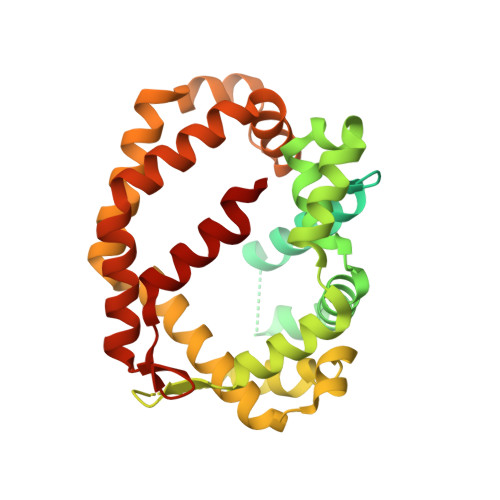
Structure of a lipid-bound viral membrane assembly protein reveals a modality for enclosing the lipid bilayer.

Pathak, P.K.Peng, S.Meng, X.Han, Y.Zhang, B.Zhang, F.Xiang, Y.Deng, J.

(2018) Proc Natl Acad Sci U S A 115: 7028-7032

  • DOI: https://doi.org/10.1073/pnas.1805855115
  • Primary Citation of Related Structures:  
    6BR8, 6BR9, 6CB6, 6CB7

  • PubMed Abstract: 

    Cellular membranes are maintained as closed compartments, broken up only transiently during membrane reorganization or lipid transportation. However, open-ended membranes, likely derived from scissions of the endoplasmic reticulum, persist in vaccinia virus-infected cells during the assembly of the viral envelope. A group of viral membrane assembly proteins (VMAPs) were identified as essential for this process. To understand the mechanism of VMAPs, we determined the 2.2-Å crystal structure of the largest member, named A6, which is a soluble protein with two distinct domains. The structure of A6 displays a novel protein fold composed mainly of alpha helices. The larger C-terminal domain forms a unique cage that encloses multiple glycerophospholipids with a lipid bilayer-like configuration. The smaller N-terminal domain does not bind lipid but negatively affects lipid binding by A6. Mutations of key hydrophobic residues lining the lipid-binding cage disrupt lipid binding and abolish viral replication. Our results reveal a protein modality for enclosing the lipid bilayer and provide molecular insight into a viral machinery involved in generating and/or stabilizing open-ended membranes.


  • Organizational Affiliation

    Department of Biochemistry and Molecular Biology, Oklahoma State University, Stillwater, OK 74078.


Macromolecules
Find similar proteins by:  (by identity cutoff)  |  3D Structure
Entity ID: 1
MoleculeChains Sequence LengthOrganismDetailsImage
Protein A6 homolog359Fowlpox virus strain NVSLMutation(s): 0 
Gene Names: FPV170
UniProt
Find proteins for Q9J563 (Fowlpox virus (strain NVSL))
Explore Q9J563 
Go to UniProtKB:  Q9J563
Entity Groups  
Sequence Clusters30% Identity50% Identity70% Identity90% Identity95% Identity100% Identity
UniProt GroupQ9J563
Sequence Annotations
Expand
  • Reference Sequence
Small Molecules
Ligands 2 Unique
IDChains Name / Formula / InChI Key2D Diagram3D Interactions
PGV
Query on PGV

Download Ideal Coordinates CCD File 
E [auth A](1R)-2-{[{[(2S)-2,3-DIHYDROXYPROPYL]OXY}(HYDROXY)PHOSPHORYL]OXY}-1-[(PALMITOYLOXY)METHYL]ETHYL (11E)-OCTADEC-11-ENOATE
C40 H77 O10 P
ADYWCMPUNIVOEA-GPJPVTGXSA-N
6OU
Query on 6OU

Download Ideal Coordinates CCD File 
B [auth A],
C [auth A],
D [auth A],
F [auth A]
[(2~{R})-1-[2-azanylethoxy(oxidanyl)phosphoryl]oxy-3-hexadecanoyloxy-propan-2-yl] (~{Z})-octadec-9-enoate
C39 H76 N O8 P
FHQVHHIBKUMWTI-OTMQOFQLSA-N
Experimental Data & Validation

Experimental Data

  • Method: X-RAY DIFFRACTION
  • Resolution: 2.20 Å
  • R-Value Free: 0.270 
  • R-Value Work: 0.216 
  • R-Value Observed: 0.221 
  • Space Group: C 2 2 21
Unit Cell:
Length ( Å )Angle ( ˚ )
a = 54.699α = 90
b = 74.151β = 90
c = 143.571γ = 90
Software Package:
Software NamePurpose
PHENIXrefinement
HKL-3000data reduction
HKL-3000data scaling
PHENIXphasing

Structure Validation

View Full Validation Report


Ligand Structure Quality Assessment 


Entry History & Funding Information

Deposition Data


Funding OrganizationLocationGrant Number
National Institutes of Health/National Institute Of Allergy and Infectious Diseases (NIH/NIAID)United StatesAI 133589

Revision History  (Full details and data files)

  • Version 1.0: 2018-06-20
    Type: Initial release
  • Version 1.1: 2018-07-04
    Changes: Data collection, Database references, Source and taxonomy
  • Version 1.2: 2018-07-18
    Changes: Data collection, Database references
  • Version 1.3: 2019-02-20
    Changes: Author supporting evidence, Data collection
  • Version 1.4: 2019-12-11
    Changes: Author supporting evidence
  • Version 1.5: 2024-03-13
    Changes: Advisory, Data collection, Database references, Structure summary