6BQN

Cryo-EM structure of ENaC


Experimental Data Snapshot

  • Method: ELECTRON MICROSCOPY
  • Resolution: 3.90 Å
  • Aggregation State: PARTICLE 
  • Reconstruction Method: SINGLE PARTICLE 

wwPDB Validation   3D Report Full Report


This is version 1.2 of the entry. See complete history


Literature

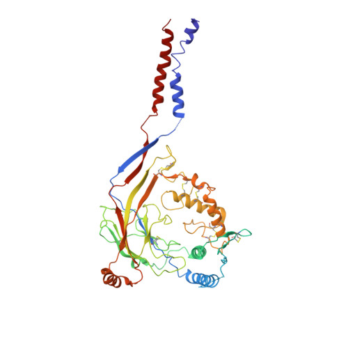
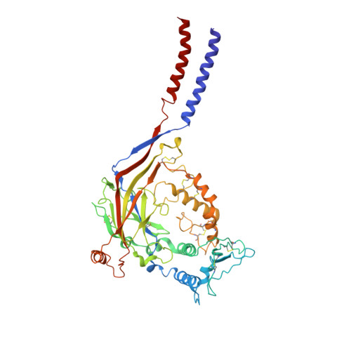
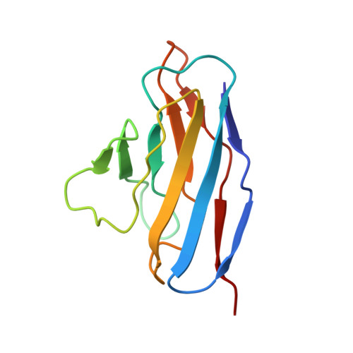
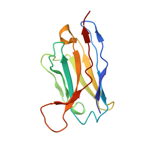
Structure of the human epithelial sodium channel by cryo-electron microscopy.

Noreng, S.Bharadwaj, A.Posert, R.Yoshioka, C.Baconguis, I.

(2018) Elife 7

  • DOI: https://doi.org/10.7554/eLife.39340
  • Primary Citation of Related Structures:  
    6BQN

  • PubMed Abstract: 

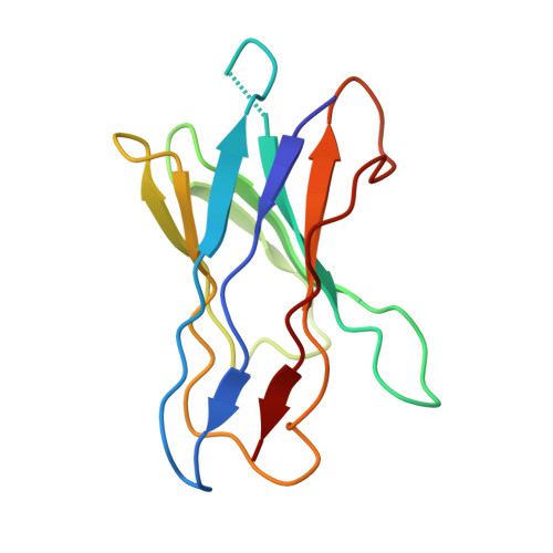
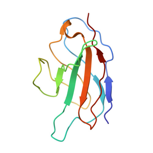
    The epithelial sodium channel (ENaC), a member of the ENaC/DEG superfamily, regulates Na + and water homeostasis. ENaCs assemble as heterotrimeric channels that harbor protease-sensitive domains critical for gating the channel. Here, we present the structure of human ENaC in the uncleaved state determined by single-particle cryo-electron microscopy. The ion channel is composed of a large extracellular domain and a narrow transmembrane domain. The structure reveals that ENaC assembles with a 1:1:1 stoichiometry of α:β:γ subunits arranged in a counter-clockwise manner. The shape of each subunit is reminiscent of a hand with key gating domains of a 'finger' and a 'thumb.' Wedged between these domains is the elusive protease-sensitive inhibitory domain poised to regulate conformational changes of the 'finger' and 'thumb'; thus, the structure provides the first view of the architecture of inhibition of ENaC.


  • Organizational Affiliation

    Department of Biochemistry & Molecular Biology, Oregon Health and Science University, Portland, United States.


Macromolecules
Find similar proteins by:  (by identity cutoff)  |  3D Structure
Entity ID: 1
MoleculeChains Sequence LengthOrganismDetailsImage
SCNN1A489Homo sapiensMutation(s): 0 
Membrane Entity: Yes 
UniProt & NIH Common Fund Data Resources
Find proteins for P37088 (Homo sapiens)
Explore P37088 
Go to UniProtKB:  P37088
PHAROS:  P37088
GTEx:  ENSG00000111319 
Entity Groups  
Sequence Clusters30% Identity50% Identity70% Identity90% Identity95% Identity100% Identity
UniProt GroupP37088
Sequence Annotations
Expand
  • Reference Sequence
Find similar proteins by:  (by identity cutoff)  |  3D Structure
Entity ID: 2
MoleculeChains Sequence LengthOrganismDetailsImage
SCNN1B495Homo sapiensMutation(s): 0 
Membrane Entity: Yes 
UniProt & NIH Common Fund Data Resources
Find proteins for P51168 (Homo sapiens)
Explore P51168 
Go to UniProtKB:  P51168
PHAROS:  P51168
GTEx:  ENSG00000168447 
Entity Groups  
Sequence Clusters30% Identity50% Identity70% Identity90% Identity95% Identity100% Identity
UniProt GroupP51168
Sequence Annotations
Expand
  • Reference Sequence
Find similar proteins by:  (by identity cutoff)  |  3D Structure
Entity ID: 3
MoleculeChains Sequence LengthOrganismDetailsImage
EGFP-SCNN1G chimera503Homo sapiensMutation(s): 0 
Membrane Entity: Yes 
UniProt & NIH Common Fund Data Resources
Find proteins for P51170 (Homo sapiens)
Explore P51170 
Go to UniProtKB:  P51170
PHAROS:  P51170
GTEx:  ENSG00000166828 
Entity Groups  
Sequence Clusters30% Identity50% Identity70% Identity90% Identity95% Identity100% Identity
UniProt GroupP51170
Sequence Annotations
Expand
  • Reference Sequence
Find similar proteins by:  (by identity cutoff)  |  3D Structure
Entity ID: 4
MoleculeChains Sequence LengthOrganismDetailsImage
7B1 fab106Mus musculusMutation(s): 0 
Sequence Annotations
Expand
  • Reference Sequence
Find similar proteins by:  (by identity cutoff)  |  3D Structure
Entity ID: 5
MoleculeChains Sequence LengthOrganismDetailsImage
7B1 fab121Mus musculusMutation(s): 0 
Sequence Annotations
Expand
  • Reference Sequence
Find similar proteins by:  (by identity cutoff)  |  3D Structure
Entity ID: 6
MoleculeChains Sequence LengthOrganismDetailsImage
10D4 fab115Mus musculusMutation(s): 0 
Sequence Annotations
Expand
  • Reference Sequence
Find similar proteins by:  (by identity cutoff)  |  3D Structure
Entity ID: 7
MoleculeChains Sequence LengthOrganismDetailsImage
10D4 fab112Mus musculusMutation(s): 0 
Sequence Annotations
Expand
  • Reference Sequence
Experimental Data & Validation

Experimental Data

  • Method: ELECTRON MICROSCOPY
  • Resolution: 3.90 Å
  • Aggregation State: PARTICLE 
  • Reconstruction Method: SINGLE PARTICLE 

Structure Validation

View Full Validation Report


Entry History & Funding Information

Deposition Data


Funding OrganizationLocationGrant Number
National Institutes of Health/Office of the DirectorUnited States--

Revision History  (Full details and data files)

  • Version 1.0: 2018-10-10
    Type: Initial release
  • Version 1.1: 2018-10-24
    Changes: Data collection, Database references, Structure summary
  • Version 1.2: 2019-12-18
    Changes: Author supporting evidence, Other