6BNX

Crystal structure of V278E-glyoxalase I mutant from Zea mays in space group P6(3)


Experimental Data Snapshot

  • Method: X-RAY DIFFRACTION
  • Resolution: 1.80 Å
  • R-Value Free: 0.224 
  • R-Value Work: 0.183 
  • R-Value Observed: 0.185 

wwPDB Validation   3D Report Full Report


This is version 1.3 of the entry. See complete history


Literature

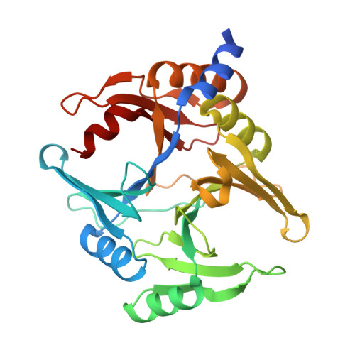
Deciphering the number and location of active sites in the monomeric glyoxalase I of Zea mays.

Gonzalez, J.M.Agostini, R.B.Alvarez, C.E.Klinke, S.Andreo, C.S.Campos-Bermudez, V.A.

(2019) FEBS J 286: 3255-3271

  • DOI: https://doi.org/10.1111/febs.14855
  • Primary Citation of Related Structures:  
    6BNN, 6BNX, 6BNZ

  • PubMed Abstract: 

    Detoxification of methylglyoxal, a toxic by-product of central sugar metabolism, is a major issue for all forms of life. The glyoxalase pathway evolved to effectively convert methylglyoxal into d-lactate via a glutathione hemithioacetal intermediate. Recently, we have shown that the monomeric glyoxalase I from maize exhibits a symmetric fold with two cavities, potentially harboring two active sites, in analogy with homodimeric enzyme surrogates. Here we confirm that only one of the two cavities exhibits glyoxalase I activity and show that it adopts a tunnel-shaped structure upon substrate binding. Such conformational change gives rise to independent binding sites for glutathione and methylglyoxal in the same active site, with important implications for the molecular reaction mechanism, which has been a matter of debate for several decades. DATABASE: Structural data are available in The Protein Data Bank database under the accession numbers 6BNN, 6BNX, and 6BNZ.


  • Organizational Affiliation

    Instituto de Bionanotecnología del NOA (INBIONATEC-CONICET), Universidad Nacional de Santiago del Estero (UNSE), Argentina.


Macromolecules
Find similar proteins by:  (by identity cutoff)  |  3D Structure
Entity ID: 1
MoleculeChains Sequence LengthOrganismDetailsImage
Lactoylglutathione lyase296Zea maysMutation(s): 1 
EC: 4.4.1.5
UniProt
Find proteins for B6TPH0 (Zea mays)
Explore B6TPH0 
Go to UniProtKB:  B6TPH0
Entity Groups  
Sequence Clusters30% Identity50% Identity70% Identity90% Identity95% Identity100% Identity
UniProt GroupB6TPH0
Sequence Annotations
Expand
  • Reference Sequence
Small Molecules
Ligands 1 Unique
IDChains Name / Formula / InChI Key2D Diagram3D Interactions
CO (Subject of Investigation/LOI)
Query on CO

Download Ideal Coordinates CCD File 
B [auth A]COBALT (II) ION
Co
XLJKHNWPARRRJB-UHFFFAOYSA-N
Experimental Data & Validation

Experimental Data

  • Method: X-RAY DIFFRACTION
  • Resolution: 1.80 Å
  • R-Value Free: 0.224 
  • R-Value Work: 0.183 
  • R-Value Observed: 0.185 
  • Space Group: P 63
Unit Cell:
Length ( Å )Angle ( ˚ )
a = 82.11α = 90
b = 82.11β = 90
c = 74.97γ = 120
Software Package:
Software NamePurpose
REFMACrefinement
XDSdata reduction
XDSdata scaling
PHASERphasing

Structure Validation

View Full Validation Report


Entry History 

Deposition Data

Revision History  (Full details and data files)

  • Version 1.0: 2018-11-21
    Type: Initial release
  • Version 1.1: 2019-06-05
    Changes: Data collection, Database references
  • Version 1.2: 2019-09-04
    Changes: Data collection, Database references
  • Version 1.3: 2023-10-04
    Changes: Data collection, Database references, Refinement description