A Novel Polar Core and Weakly Fixed C-Tail in Squid Arrestin Provide New Insight into Interaction with Rhodopsin.
Bandyopadhyay, A., Van Eps, N., Eger, B.T., Rauscher, S., Yedidi, R.S., Moroni, T., West, G.M., Robinson, K.A., Griffin, P.R., Mitchell, J., Ernst, O.P.(2018) J Mol Biol 430: 4102-4118
- PubMed: 30120952 Search on PubMed
- DOI: https://doi.org/10.1016/j.jmb.2018.08.009
- Primary Citation Related Structures:
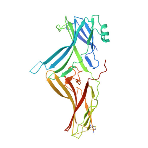
6BK9 - PubMed Abstract:
Photoreceptors of the squid Loligo pealei contain a G-protein-coupled receptor (GPCR) signaling system that activates phospholipase C in response to light. Analogous to the mammalian visual system, signaling of the photoactivated GPCR rhodopsin is terminated by binding of squid arrestin (sArr). sArr forms a light-dependent, high-affinity complex with squid rhodopsin, which does not require prior receptor phosphorylation for interaction. This is at odds with classical mammalian GPCR desensitization where an agonist-bound phosphorylated receptor is needed to break stabilizing constraints within arrestins, the so-called "three-element interaction" and "polar core" network, before a stable receptor-arrestin complex can be established. Biophysical and mass spectrometric analysis of the squid rhodopsin-arrestin complex indicates that in contrast to mammalian arrestins, the sArr C-tail is not involved in a stable three-element interaction. We determined the crystal structure of C-terminally truncated sArr that adopts a basal conformation common to arrestins and is stabilized by a series of weak but novel polar core interactions. Unlike mammalian arrestin-1, deletion of the sArr C-tail does not influence kinetic properties of complex formation of sArr with the receptor. Hydrogen-deuterium exchange studies revealed the footprint of the light-activated rhodopsin on sArr. Furthermore, double electron-electron resonance spectroscopy experiments provide evidence that receptor-bound sArr adopts a conformation different from the one known for arrestin-1 and molecular dynamics simulations reveal the residues that account for the weak three-element interaction. Insights gleaned from studying this system add to our general understanding of GPCR-arrestin interaction.
- Department of Biochemistry, University of Toronto, Toronto, Ontario M5S 1A8, Canada.
Organizational Affiliation: