6BHX

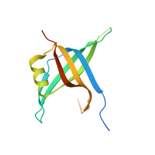
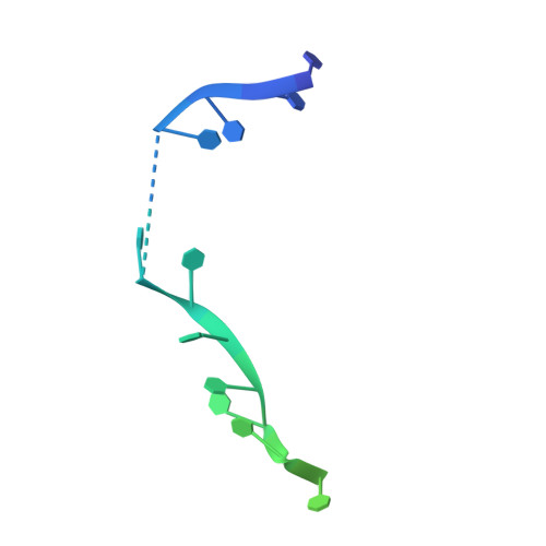
B. subtilis SsbA with DNA


Experimental Data Snapshot

  • Method: X-RAY DIFFRACTION
  • Resolution: 2.94 Å
  • R-Value Free: 0.269 
  • R-Value Work: 0.216 
  • R-Value Observed: 0.219 

wwPDB Validation   3D Report Full Report


This is version 1.4 of the entry. See complete history


Literature

Structural Mechanisms of Cooperative DNA Binding by Bacterial Single-Stranded DNA-Binding Proteins.

Dubiel, K.Myers, A.R.Kozlov, A.G.Yang, O.Zhang, J.Ha, T.Lohman, T.M.Keck, J.L.

(2019) J Mol Biol 431: 178-195

  • DOI: https://doi.org/10.1016/j.jmb.2018.11.019
  • Primary Citation of Related Structures:  
    6BHW, 6BHX

  • PubMed Abstract: 

    Bacteria encode homooligomeric single-stranded (ss) DNA-binding proteins (SSBs) that coat and protect ssDNA intermediates formed during genome maintenance reactions. The prototypical Escherichia coli SSB tetramer can bind ssDNA using multiple modes that differ by the number of bases bound per tetramer and the magnitude of the binding cooperativity. Our understanding of the mechanisms underlying cooperative ssDNA binding by SSBs has been hampered by the limited amount of structural information available for interfaces that link adjacent SSB proteins on ssDNA. Here we present a crystal structure of Bacillus subtilis SsbA bound to ssDNA. The structure resolves SsbA tetramers joined together by a ssDNA "bridge" and identifies an interface, termed the "bridge interface," that links adjacent SSB tetramers through an evolutionarily conserved surface near the ssDNA-binding site. E. coli SSB variants with altered bridge interface residues bind ssDNA with reduced cooperativity and with an altered distribution of DNA binding modes. These variants are also more readily displaced from ssDNA by RecA than wild-type SSB. In spite of these biochemical differences, each variant is able to complement deletion of the ssb gene in E. coli. Together our data suggest a model in which the bridge interface contributes to cooperative ssDNA binding and SSB function but that destabilization of the bridge interface is tolerated in cells.


  • Organizational Affiliation

    Department of Biomolecular Chemistry, University of Wisconsin School of Medicine and Public Health, Madison, WI 53706, USA.


Macromolecules

Find similar proteins by:  (by identity cutoff)  |  3D Structure
Entity ID: 1
MoleculeChains Sequence LengthOrganismDetailsImage
Single-stranded DNA-binding protein A
A, B, C, D
132Bacillus subtilis subsp. subtilis str. 168Mutation(s): 0 
Gene Names: ssbABSU40900
UniProt
Find proteins for P37455 (Bacillus subtilis (strain 168))
Explore P37455 
Go to UniProtKB:  P37455
Entity Groups  
Sequence Clusters30% Identity50% Identity70% Identity90% Identity95% Identity100% Identity
UniProt GroupP37455
Sequence Annotations
Expand
  • Reference Sequence
Find similar nucleic acids by:  (by identity cutoff)  |  3D Structure
Entity ID: 2
MoleculeChains LengthOrganismImage
DNA (5'-D(P*TP*TP*TP*TP*TP*TP*TP*TP*TP*TP*T)-3')30synthetic construct
Sequence Annotations
Expand
  • Reference Sequence
Experimental Data & Validation

Experimental Data

  • Method: X-RAY DIFFRACTION
  • Resolution: 2.94 Å
  • R-Value Free: 0.269 
  • R-Value Work: 0.216 
  • R-Value Observed: 0.219 
  • Space Group: P 21 21 21
Unit Cell:
Length ( Å )Angle ( ˚ )
a = 57.939α = 90
b = 93.681β = 90
c = 100.968γ = 90
Software Package:
Software NamePurpose
HKL-2000data processing
PHENIXrefinement
ADSCdata collection
HKL-2000data reduction
HKL-2000data scaling
Cootmodel building
PHENIXphasing

Structure Validation

View Full Validation Report


Entry History & Funding Information

Deposition Data


Funding OrganizationLocationGrant Number
National Institutes of Health/National Institute of General Medical Sciences (NIH/NIGMS)United StatesR01 GM098885

Revision History  (Full details and data files)

  • Version 1.0: 2018-12-19
    Type: Initial release
  • Version 1.1: 2019-01-30
    Changes: Data collection, Database references
  • Version 1.2: 2019-02-20
    Changes: Author supporting evidence, Data collection
  • Version 1.3: 2020-01-01
    Changes: Author supporting evidence
  • Version 1.4: 2024-03-13
    Changes: Data collection, Database references