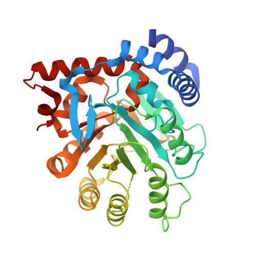
Crystal structure of dihydroorotate dehydrogenase from Helicobacter pylori with bound flavin mononucleotide.
Agarwal, A.A., Georgiades, J.D., Dranow, D.M., Lorimer, D.D., Edwards, T., Barrett, K.F., Craig, J.K., Van Voorhis, W.C., Myler, P.J., Smith, C.L.(2025) Acta Crystallogr F Struct Biol Commun 81: 108-117
- PubMed: 39960828 Search on PubMedSearch on PubMed Central
- DOI: https://doi.org/10.1107/S2053230X25000858
- Primary Citation Related Structures:
6B8S - PubMed Abstract:
Helicobacter pylori is the primary causative agent of peptic ulcer disease, among other gastrointestinal ailments, and currently affects over half of the global population. Although some treatments exist, growing resistance to these drugs has prompted efforts to develop novel approaches to fighting this pathogen. To generate many of the nucleotides essential to biochemical processes, H. pylori relies exclusively on the de novo biosynthesis of these molecules. Recent drug-discovery efforts have targeted the first committed step of this pathway, catalysed by a class 2 dihydroorotate dehydrogenase (DHODH). However, these initiatives have been limited by the lack of a crystal structure. Here, we detail the crystal structure of H. pylori DHODH (HpDHODH) at 2.25 Å resolution (PDB entry 6b8s). We performed a large-scale bioinformatics search to find evolutionary homologs. Our results indicate that HpDHODH shows high conservation of both sequence and structure in its active site. We identified key polar interactions between the HpDHODH protein and its requisite flavin mononucleotide (FMN) cofactor, identifying amino-acid residues that are critical to its function. Most notably, we found that HpDHODH maintains several structural features that allow it to associate with the inner membrane and utilize ubiquinone to achieve catalytic turnover. We discovered a hydrophobic channel that runs from the putative membrane interface on the N-terminal microdomain to the core of the protein. We predict that this channel establishes a connection between the ubiquinone pool in the membrane and the FMN in the active site. These findings provide a structural explanation for the competitive inhibition of ubiquinone by pyrazole-based compounds that was determined biochemically in other studies. Understanding this mechanism may facilitate the development of new drugs targeting this enzyme and push the effort to find a resistance-free treatment for H. pylori.
- Department of Biology, Washington University in St Louis School of Medicine, St Louis, MO 63114, USA.
Organizational Affiliation: