6B8O | pdb_00006b8o

WT Ig-like V Domain with Phosphatidylserine


Experimental Data Snapshot

  • Method: X-RAY DIFFRACTION
  • Resolution: 2.20 Å
  • R-Value Free: 
    0.261 (Depositor), 0.265 (DCC) 
  • R-Value Work: 
    0.222 (Depositor), 0.228 (DCC) 
  • R-Value Observed: 
    0.224 (Depositor) 

Starting Model: experimental
View more details

wwPDB Validation 3D Report Full Report

Validation slider image for 6B8O

Ligand Structure Quality Assessment 


This is version 1.4 of the entry. See complete history

Literature

Molecular basis for the loss-of-function effects of the Alzheimer's disease-associated R47H variant of the immune receptor TREM2.

Sudom, A.Talreja, S.Danao, J.Bragg, E.Kegel, R.Min, X.Richardson, J.Zhang, Z.Sharkov, N.Marcora, E.Thibault, S.Bradley, J.Wood, S.Lim, A.C.Chen, H.Wang, S.Foltz, I.N.Sambashivan, S.Wang, Z.

(2018) J Biological Chem 293: 12634-12646

  • DOI: https://doi.org/10.1074/jbc.RA118.002352
  • Primary Citation Related Structures: 
    5UD7, 5UD8, 6B8O

  • PubMed Abstract: 

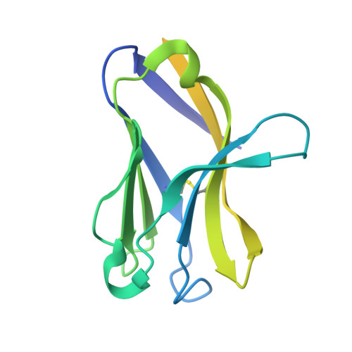
    Triggering receptor expressed on myeloid cells 2 (TREM2) is an immune receptor expressed on the surface of microglia, macrophages, dendritic cells, and osteoclasts. The R47H TREM2 variant is a significant risk factor for late-onset Alzheimer's disease (AD), and the molecular basis of R47H TREM2 loss of function is an emerging area of TREM2 biology. Here, we report three high-resolution structures of the extracellular ligand-binding domains (ECDs) of R47H TREM2, apo-WT, and phosphatidylserine (PS)-bound WT TREM2 at 1.8, 2.2, and 2.2 Å, respectively. The structures reveal that Arg 47 plays a critical role in maintaining the structural features of the complementarity-determining region 2 (CDR2) loop and the putative positive ligand-interacting surface (PLIS), stabilizing conformations capable of ligand interaction. This is exemplified in the PS-bound structure, in which the CDR2 loop and PLIS drive critical interactions with PS via surfaces that are disrupted in the variant. Together with in vitro and in vivo characterization, our structural findings elucidate the molecular mechanism underlying loss of ligand binding, putative oligomerization, and functional activity of R47H TREM2. They also help unravel how decreased in vitro and in vivo stability of TREM2 contribute to loss of function in disease.


  • Organizational Affiliation
    • From Amgen Discovery Research, Amgen Inc., San Francisco, California 94080, asudom@amgen.com.

Macromolecule Content 

  • Total Structure Weight: 120.09 kDa 
  • Atom Count: 5,416 
  • Modeled Residue Count: 643 
  • Deposited Residue Count: 1,014 
  • Unique protein chains: 1

Macromolecules

Find similar proteins by:|  3D Structure
Entity ID: 1
MoleculeChains  Sequence LengthOrganismDetailsImage
Triggering receptor expressed on myeloid cells 2
A, B, C, D, E
A, B, C, D, E, F
169Homo sapiensMutation(s): 0 
Gene Names: TREM2
UniProt & NIH Common Fund Data Resources
Find proteins for Q9NZC2 (Homo sapiens)
Explore Q9NZC2 
Go to UniProtKB:  Q9NZC2
PHAROS:  Q9NZC2
GTEx:  ENSG00000095970 
Entity Groups
Sequence Clusters30% Identity50% Identity70% Identity90% Identity95% Identity100% Identity
UniProt GroupQ9NZC2
Glycosylation
Glycosylation Sites: 1Go to GlyGen: Q9NZC2-1
Sequence Annotations
Expand
Reference Sequence

Small Molecules

Ligands 5 Unique
IDChains Name / Formula / InChI Key2D Diagram3D Interactions
PSF

Query on PSF


Download:Ideal Coordinates CCD File
BA [auth C],
GA [auth D],
U [auth B]
1,2-DICAPROYL-SN-PHOSPHATIDYL-L-SERINE
C18 H34 N O10 P
MIQYPPGTNIFAPO-CABCVRRESA-N
NAG

Query on NAG


Download:Ideal Coordinates CCD File
CA [auth D]
G [auth A]
HA [auth E]
MA [auth F]
R [auth B]
CA [auth D],
G [auth A],
HA [auth E],
MA [auth F],
R [auth B],
Z [auth C]
2-acetamido-2-deoxy-beta-D-glucopyranose
C8 H15 N O6
OVRNDRQMDRJTHS-FMDGEEDCSA-N
IOD

Query on IOD


Download:Ideal Coordinates CCD File
AA [auth C]
H [auth A]
I [auth A]
IA [auth E]
J [auth A]
AA [auth C],
H [auth A],
I [auth A],
IA [auth E],
J [auth A],
S [auth B],
T [auth B]
IODIDE ION
I
XMBWDFGMSWQBCA-UHFFFAOYSA-M
SO4

Query on SO4


Download:Ideal Coordinates CCD File
DA [auth D]
EA [auth D]
FA [auth D]
JA [auth E]
K [auth A]
DA [auth D],
EA [auth D],
FA [auth D],
JA [auth E],
K [auth A],
KA [auth E],
L [auth A],
LA [auth E],
NA [auth F],
OA [auth F],
PA [auth F]
SULFATE ION
O4 S
QAOWNCQODCNURD-UHFFFAOYSA-L
EDO

Query on EDO


Download:Ideal Coordinates CCD File
M [auth A]
N [auth A]
O [auth A]
P [auth A]
Q [auth A]
M [auth A],
N [auth A],
O [auth A],
P [auth A],
Q [auth A],
QA [auth F],
V [auth B],
W [auth B],
X [auth B],
Y [auth B]
1,2-ETHANEDIOL
C2 H6 O2
LYCAIKOWRPUZTN-UHFFFAOYSA-N

Experimental Data & Validation

Experimental Data

  • Method: X-RAY DIFFRACTION
  • Resolution: 2.20 Å
  • R-Value Free:  0.261 (Depositor), 0.265 (DCC) 
  • R-Value Work:  0.222 (Depositor), 0.228 (DCC) 
  • R-Value Observed: 0.224 (Depositor) 
Space Group: P 41 21 2
Unit Cell:
Length ( Å )Angle ( ˚ )
a = 160.331α = 90
b = 160.331β = 90
c = 86.205γ = 90
Software Package:
Software NamePurpose
REFMACrefinement
Aimlessdata scaling
PDB_EXTRACTdata extraction

Structure Validation

View Full Validation Report


Ligand Structure Quality Assessment 


Entry History 

Deposition Data

Revision History  (Full details and data files)

  • Version 1.0: 2018-06-27
    Type: Initial release
  • Version 1.1: 2018-08-22
    Changes: Data collection, Database references
  • Version 1.2: 2020-07-29
    Type: Remediation
    Reason: Carbohydrate remediation
    Changes: Data collection, Derived calculations, Structure summary
  • Version 1.3: 2023-10-04
    Changes: Data collection, Database references, Refinement description, Structure summary
  • Version 1.4: 2024-11-13
    Changes: Structure summary