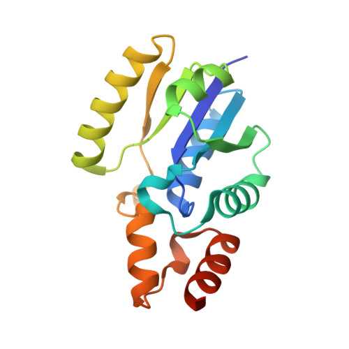
6B7A

Crystal structure of E.coli Phosphopantetheine Adenylyltransferase (PPAT/CoaD) in complex with 2-methyl-1H-benzo[d]imidazol-4-ol


Experimental Data Snapshot

  • Method: X-RAY DIFFRACTION
  • Resolution: 1.99 Å
  • R-Value Free: 0.199 
  • R-Value Work: 0.174 
  • R-Value Observed: 0.175 

wwPDB Validation   3D Report Full Report


Ligand Structure Quality Assessment 


This is version 1.1 of the entry. See complete history


Literature

High-Confidence Protein-Ligand Complex Modeling by NMR-Guided Docking Enables Early Hit Optimization.

Proudfoot, A.Bussiere, D.E.Lingel, A.

(2017) J Am Chem Soc 139: 17824-17833

  • DOI: https://doi.org/10.1021/jacs.7b07171
  • Primary Citation of Related Structures:  
    6B7A, 6B7B, 6B7C, 6B7D, 6B7E, 6B7F

  • PubMed Abstract: 

    Structure-based drug design is an integral part of modern day drug discovery and requires detailed structural characterization of protein-ligand interactions, which is most commonly performed by X-ray crystallography. However, the success rate of generating these costructures is often variable, in particular when working with dynamic proteins or weakly binding ligands. As a result, structural information is not routinely obtained in these scenarios, and ligand optimization is challenging or not pursued at all, representing a substantial limitation in chemical scaffolds and diversity. To overcome this impediment, we have developed a robust NMR restraint guided docking protocol to generate high-quality models of protein-ligand complexes. By combining the use of highly methyl-labeled protein with experimentally determined intermolecular distances, a comprehensive set of protein-ligand distances is generated which then drives the docking process and enables the determination of the correct ligand conformation in the bound state. For the first time, the utility and performance of such a method is fully demonstrated by employing the generated models for the successful, prospective optimization of crystallographically intractable fragment hits into more potent binders.


  • Organizational Affiliation

    Global Discovery Chemistry, Novartis Institutes for BioMedical Research , 5300 Chiron Way, Emeryville, California 94608, United States.


Macromolecules
Find similar proteins by:  (by identity cutoff)  |  3D Structure
Entity ID: 1
MoleculeChains Sequence LengthOrganismDetailsImage
Phosphopantetheine adenylyltransferase
A, B
167Escherichia coli K-12Mutation(s): 0 
Gene Names: coaDkdtByicAb3634JW3609
EC: 2.7.7.3
UniProt
Find proteins for P0A6I6 (Escherichia coli (strain K12))
Explore P0A6I6 
Go to UniProtKB:  P0A6I6
Entity Groups  
Sequence Clusters30% Identity50% Identity70% Identity90% Identity95% Identity100% Identity
UniProt GroupP0A6I6
Sequence Annotations
Expand
  • Reference Sequence
Small Molecules
Ligands 5 Unique
IDChains Name / Formula / InChI Key2D Diagram3D Interactions
POP
Query on POP

Download Ideal Coordinates CCD File 
N [auth B]PYROPHOSPHATE 2-
H2 O7 P2
XPPKVPWEQAFLFU-UHFFFAOYSA-L
CWM (Subject of Investigation/LOI)
Query on CWM

Download Ideal Coordinates CCD File 
C [auth A],
I [auth B]
2-methyl-1H-benzimidazol-7-ol
C8 H8 N2 O
HLPAESMITTURFN-UHFFFAOYSA-N
PEG
Query on PEG

Download Ideal Coordinates CCD File 
F [auth A],
G [auth A],
M [auth B]
DI(HYDROXYETHYL)ETHER
C4 H10 O3
MTHSVFCYNBDYFN-UHFFFAOYSA-N
SO4
Query on SO4

Download Ideal Coordinates CCD File 
D [auth A],
E [auth A],
J [auth B],
K [auth B],
L [auth B]
SULFATE ION
O4 S
QAOWNCQODCNURD-UHFFFAOYSA-L
CL
Query on CL

Download Ideal Coordinates CCD File 
H [auth A]CHLORIDE ION
Cl
VEXZGXHMUGYJMC-UHFFFAOYSA-M
Binding Affinity Annotations 
IDSourceBinding Affinity
CWM Binding MOAD:  6B7A Kd: 3.20e+4 (nM) from 1 assay(s)
Experimental Data & Validation

Experimental Data

  • Method: X-RAY DIFFRACTION
  • Resolution: 1.99 Å
  • R-Value Free: 0.199 
  • R-Value Work: 0.174 
  • R-Value Observed: 0.175 
  • Space Group: I 2 3
Unit Cell:
Length ( Å )Angle ( ˚ )
a = 134.6α = 90
b = 134.6β = 90
c = 134.6γ = 90
Software Package:
Software NamePurpose
Aimlessdata scaling
PHENIXrefinement
PDB_EXTRACTdata extraction
iMOSFLMdata reduction
PHENIXphasing
Cootmodel building

Structure Validation

View Full Validation Report


Ligand Structure Quality Assessment 


Entry History 

Deposition Data

Revision History  (Full details and data files)

  • Version 1.0: 2017-12-27
    Type: Initial release
  • Version 1.1: 2023-10-04
    Changes: Data collection, Database references, Refinement description