6B5C

Structural Basis for Katanin Self-Assembly


Experimental Data Snapshot

  • Method: X-RAY DIFFRACTION
  • Resolution: 2.40 Å
  • R-Value Free: 0.253 
  • R-Value Work: 0.215 
  • R-Value Observed: 0.217 

wwPDB Validation   3D Report Full Report


This is version 1.3 of the entry. See complete history


Literature

Structural basis for disassembly of katanin heterododecamers.

Nithianantham, S.McNally, F.J.Al-Bassam, J.

(2018) J Biol Chem 293: 10590-10605

  • DOI: https://doi.org/10.1074/jbc.RA117.001215
  • Primary Citation of Related Structures:  
    6B5C, 6B5D

  • PubMed Abstract: 

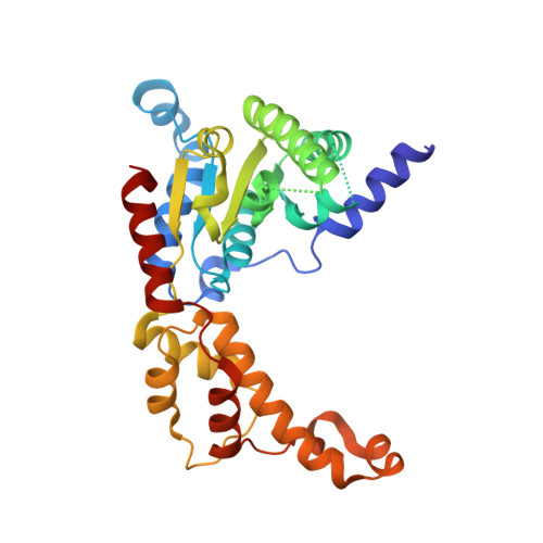
    The reorganization of microtubules in mitosis, meiosis, and development requires the microtubule-severing activity of katanin. Katanin is a heterodimer composed of an A TPase a ssociated with diverse cellular a ctivities (AAA) subunit and a regulatory subunit. Microtubule severing requires ATP hydrolysis by katanin's conserved AAA ATPase domains. Whereas other AAA ATPases form stable hexamers, we show that katanin forms only a monomer or dimers of heterodimers in solution. Katanin oligomers consistent with hexamers of heterodimers or heterododecamers were only observed for an ATP hydrolysis-deficient mutant in the presence of ATP. X-ray structures of katanin's AAA ATPase in monomeric nucleotide-free and pseudo-oligomeric ADP-bound states revealed conformational changes in the AAA subdomains that explained the structural basis for the instability of the katanin heterododecamer. We propose that the rapid dissociation of katanin AAA oligomers may lead to an autoinhibited state that prevents inappropriate microtubule severing or that cyclical disassembly into heterodimers may critically contribute to the microtubule-severing mechanism.


  • Organizational Affiliation

    From the Department of Molecular Cellular Biology University of California, Davis, California 95616.


Macromolecules
Find similar proteins by:  (by identity cutoff)  |  3D Structure
Entity ID: 1
MoleculeChains Sequence LengthOrganismDetailsImage
Katanin p60 ATPase-containing subunit A-like 1307Homo sapiensMutation(s): 1 
Gene Names: KATNAL1
EC: 3.6.4.3
UniProt & NIH Common Fund Data Resources
Find proteins for Q9BW62 (Homo sapiens)
Explore Q9BW62 
Go to UniProtKB:  Q9BW62
PHAROS:  Q9BW62
GTEx:  ENSG00000102781 
Entity Groups  
Sequence Clusters30% Identity50% Identity70% Identity90% Identity95% Identity100% Identity
UniProt GroupQ9BW62
Sequence Annotations
Expand
  • Reference Sequence
Small Molecules
Ligands 1 Unique
IDChains Name / Formula / InChI Key2D Diagram3D Interactions
PG4
Query on PG4

Download Ideal Coordinates CCD File 
B [auth A]TETRAETHYLENE GLYCOL
C8 H18 O5
UWHCKJMYHZGTIT-UHFFFAOYSA-N
Experimental Data & Validation

Experimental Data

  • Method: X-RAY DIFFRACTION
  • Resolution: 2.40 Å
  • R-Value Free: 0.253 
  • R-Value Work: 0.215 
  • R-Value Observed: 0.217 
  • Space Group: P 21 21 21
Unit Cell:
Length ( Å )Angle ( ˚ )
a = 40.1α = 90
b = 61.19β = 90
c = 117.62γ = 90
Software Package:
Software NamePurpose
PHENIXrefinement
SCALAdata scaling
PHASERphasing
PDB_EXTRACTdata extraction
iMOSFLMdata reduction
Cootmodel building

Structure Validation

View Full Validation Report


Entry History & Funding Information

Deposition Data


Funding OrganizationLocationGrant Number
National Institutes of Health/National Institute of General Medical Sciences (NIH/NIGMS)United StatesGM110283

Revision History  (Full details and data files)

  • Version 1.0: 2018-05-23
    Type: Initial release
  • Version 1.1: 2018-07-18
    Changes: Data collection, Database references, Source and taxonomy
  • Version 1.2: 2020-01-01
    Changes: Author supporting evidence
  • Version 1.3: 2023-10-04
    Changes: Data collection, Database references, Refinement description