6B55 | pdb_00006b55

Crystal structure of the Plant Defensin NaD1 complexed with phosphatidic acid


Experimental Data Snapshot

  • Method: X-RAY DIFFRACTION
  • Resolution: 2.50 Å
  • R-Value Free: 
    0.252 (Depositor), 0.251 (DCC) 
  • R-Value Work: 
    0.203 (Depositor), 0.203 (DCC) 
  • R-Value Observed: 
    0.206 (Depositor) 

Starting Model: experimental
View more details

wwPDB Validation 3D Report Full Report

Validation slider image for 6B55

Ligand Structure Quality Assessment 


This is version 1.6 of the entry. See complete history

Literature

X-ray structure of a carpet-like antimicrobial defensin-phospholipid membrane disruption complex.

Jarva, M.Lay, F.T.Phan, T.K.Humble, C.Poon, I.K.H.Bleackley, M.R.Anderson, M.A.Hulett, M.D.Kvansakul, M.

(2018) Nat Commun 9: 1962-1962

  • DOI: https://doi.org/10.1038/s41467-018-04434-y
  • Primary Citation Related Structures: 
    6B55

  • PubMed Abstract: 

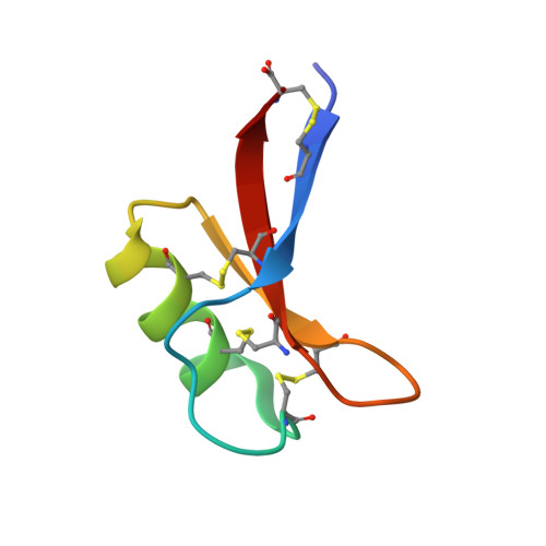
    Defensins are cationic antimicrobial peptides expressed throughout the plant and animal kingdoms as a first line of defense against pathogens. Membrane targeting and disruption is a crucial function of many defensins, however the precise mechanism remains unclear. Certain plant defensins form dimers that specifically bind the membrane phospholipids phosphatidic acid (PA) and phosphatidylinositol 4,5-bisphosphate, thereby triggering the assembly of defensin-lipid oligomers that permeabilize cell membranes. To understand this permeabilization mechanism, here we determine the crystal structure of the plant defensin NaD1 bound to PA. The structure reveals a 20-mer that adopts a concave sheet- or carpet-like topology where NaD1 dimers form one face and PA acyl chains form the other face of the sheet. Furthermore, we show that Arg39 is critical for PA binding, oligomerization and fungal cell killing. These findings identify a putative defensin-phospholipid membrane attack configuration that supports a longstanding proposed carpet mode of membrane disruption.


  • Organizational Affiliation
    • Department of Biochemistry and Genetics, La Trobe Institute for Molecular Science, La Trobe University, Melbourne, Victoria, 3086, Australia.

Macromolecule Content 

  • Total Structure Weight: 115 kDa 
  • Atom Count: 8,306 
  • Modeled Residue Count: 960 
  • Deposited Residue Count: 960 
  • Unique protein chains: 1

Macromolecules

Find similar proteins by:|  3D Structure
Entity ID: 1
MoleculeChains  Sequence LengthOrganismDetailsImage
Flower-specific defensin
A, B, C, D, E
A, B, C, D, E, F, G, H, I, J, K, L, M, N, O, P, Q, R, S, T
48Nicotiana alataMutation(s): 0 
UniProt
Find proteins for Q8GTM0 (Nicotiana alata)
Explore Q8GTM0 
Go to UniProtKB:  Q8GTM0
Entity Groups
Sequence Clusters30% Identity50% Identity70% Identity90% Identity95% Identity100% Identity
UniProt GroupQ8GTM0
Sequence Annotations
Expand
Reference Sequence

Small Molecules

Ligands 4 Unique
IDChains Name / Formula / InChI Key2D Diagram3D Interactions
PA8
(Subject of Investigation/LOI)

Query on PA8


Download:Ideal Coordinates CCD File
EA [auth H]
FA [auth I]
JA [auth L]
LA [auth M]
OA [auth P]
EA [auth H],
FA [auth I],
JA [auth L],
LA [auth M],
OA [auth P],
PA [auth Q],
RA [auth R],
SA [auth S],
TA [auth T],
U [auth A],
V [auth B],
X [auth C],
Y [auth D],
Z [auth E]
1,2-DIOCTANOYL-SN-GLYCERO-3-PHOSPHATE
C19 H36 O8 P
XYSBQYUENLDGMI-QGZVFWFLSA-M
PGE

Query on PGE


Download:Ideal Coordinates CCD File
AA [auth E],
GA [auth I],
HA [auth I],
IA [auth J],
MA [auth M]
TRIETHYLENE GLYCOL
C6 H14 O4
ZIBGPFATKBEMQZ-UHFFFAOYSA-N
PO4

Query on PO4


Download:Ideal Coordinates CCD File
BA [auth G],
CA [auth G],
W [auth B]
PHOSPHATE ION
O4 P
NBIIXXVUZAFLBC-UHFFFAOYSA-K
EDO

Query on EDO


Download:Ideal Coordinates CCD File
DA [auth G],
KA [auth L],
NA [auth O],
QA [auth Q],
UA [auth T]
1,2-ETHANEDIOL
C2 H6 O2
LYCAIKOWRPUZTN-UHFFFAOYSA-N

Experimental Data & Validation

Experimental Data

  • Method: X-RAY DIFFRACTION
  • Resolution: 2.50 Å
  • R-Value Free:  0.252 (Depositor), 0.251 (DCC) 
  • R-Value Work:  0.203 (Depositor), 0.203 (DCC) 
  • R-Value Observed: 0.206 (Depositor) 
Space Group: C 2 2 21
Unit Cell:
Length ( Å )Angle ( ˚ )
a = 114.218α = 90
b = 154.925β = 90
c = 135.999γ = 90
Software Package:
Software NamePurpose
Aimlessdata reduction
PHASERphasing
PHENIXrefinement
PDB_EXTRACTdata extraction
Cootmodel building
XDSdata processing
XDSdata scaling

Structure Validation

View Full Validation Report


Ligand Structure Quality Assessment 


Entry History 

& Funding Information

Deposition Data


Funding OrganizationLocationGrant Number
Australian Research Council (ARC)AustraliaFT130101349

Revision History  (Full details and data files)

  • Version 1.0: 2018-05-23
    Type: Initial release
  • Version 1.1: 2018-05-30
    Changes: Data collection, Database references
  • Version 1.2: 2018-08-15
    Changes: Data collection, Database references
  • Version 1.3: 2019-02-20
    Changes: Author supporting evidence, Data collection
  • Version 1.4: 2020-01-01
    Changes: Author supporting evidence
  • Version 1.5: 2023-10-04
    Changes: Data collection, Database references, Refinement description
  • Version 1.6: 2024-11-20
    Changes: Structure summary