6B2N | pdb_00006b2n

Crystal structure of TEM-1 beta-lactamase mutant M182N


Experimental Data Snapshot

  • Method: X-RAY DIFFRACTION
  • Resolution: 2.00 Å
  • R-Value Free: 
    0.283 (Depositor), 0.285 (DCC) 
  • R-Value Work: 
    0.231 (Depositor), 0.233 (DCC) 
  • R-Value Observed: 
    0.235 (Depositor) 

Starting Model: experimental
View more details

wwPDB Validation 3D Report Full Report

Validation slider image for 6B2N

This is version 1.2 of the entry. See complete history

Literature

Prediction of New Stabilizing Mutations Based on Mechanistic Insights from Markov State Models.

Zimmerman, M.I.Hart, K.M.Sibbald, C.A.Frederick, T.E.Jimah, J.R.Knoverek, C.R.Tolia, N.H.Bowman, G.R.

(2017) ACS Cent Sci 3: 1311-1321

  • DOI: https://doi.org/10.1021/acscentsci.7b00465
  • Primary Citation Related Structures: 
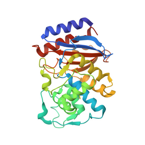
    6B2N

  • PubMed Abstract: 

    Protein stabilization is fundamental to enzyme function and evolution, yet understanding the determinants of a protein's stability remains a challenge. This is largely due to a shortage of atomically detailed models for the ensemble of relevant protein conformations and their relative populations. For example, the M182T substitution in TEM β-lactamase, an enzyme that confers antibiotic resistance to bacteria, is stabilizing but the precise mechanism remains unclear. Here, we employ Markov state models (MSMs) to uncover how M182T shifts the distribution of different structures that TEM adopts. We find that M182T stabilizes a helix that is a key component of a domain interface. We then predict the effects of other mutations, including a novel stabilizing mutation, and experimentally test our predictions using a combination of stability measurements, crystallography, NMR, and in vivo measurements of bacterial fitness. We expect our insights and methodology to provide a valuable foundation for protein design.


  • Organizational Affiliation
    • Department of Biochemistry & Molecular Biophysics, Washington University School of Medicine, 660 South Euclid Avenue, St. Louis, Missouri 63110, United States.

Macromolecule Content 

  • Total Structure Weight: 116.08 kDa 
  • Atom Count: 8,207 
  • Modeled Residue Count: 1,033 
  • Deposited Residue Count: 1,052 
  • Unique protein chains: 1

Macromolecules

Find similar proteins by:|  3D Structure
Entity ID: 1
MoleculeChains  Sequence LengthOrganismDetailsImage
Beta-lactamase TEM
A, B, C, D
263Escherichia coliMutation(s): 1 
Gene Names: blablaT-3blaT-4blaT-5blaT-6
EC: 3.5.2.6
UniProt
Find proteins for P62593 (Escherichia coli)
Explore P62593 
Go to UniProtKB:  P62593
Entity Groups
Sequence Clusters30% Identity50% Identity70% Identity90% Identity95% Identity100% Identity
UniProt GroupP62593
Sequence Annotations
Expand
Reference Sequence

Experimental Data & Validation

Experimental Data

  • Method: X-RAY DIFFRACTION
  • Resolution: 2.00 Å
  • R-Value Free:  0.283 (Depositor), 0.285 (DCC) 
  • R-Value Work:  0.231 (Depositor), 0.233 (DCC) 
  • R-Value Observed: 0.235 (Depositor) 
Space Group: P 1 21 1
Unit Cell:
Length ( Å )Angle ( ˚ )
a = 81.91α = 90
b = 49.66β = 90.1
c = 122.16γ = 90
Software Package:
Software NamePurpose
PHENIXrefinement
XDSdata reduction
XDSdata scaling
PHASERphasing

Structure Validation

View Full Validation Report


Entry History 

Deposition Data

Revision History  (Full details and data files)

  • Version 1.0: 2018-01-17
    Type: Initial release
  • Version 1.1: 2023-10-04
    Changes: Data collection, Database references, Refinement description
  • Version 1.2: 2024-10-16
    Changes: Structure summary