


Crystal structure of a nucleoside diphosphate kinase NDK from Helicobacter pylori
Edwards, T.E., Dranow, D.M., Lorimer, D.D.To be published.
Experimental Data Snapshot
Starting Model: experimental
View more details
wwPDB Validation 3D Report Full Report

Macromolecule Content
Entity ID: 1 | |||||
|---|---|---|---|---|---|
| Molecule | Chains | Sequence Length | Organism | Details | Image |
| Nucleoside diphosphate kinase | 145 | Helicobacter pylori | Mutation(s): 0 Gene Names: ndk, HPG27_182 EC: 2.7.4.6 | 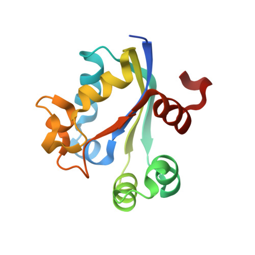 | |
UniProt | |||||
Entity Groups | |||||
| Sequence Clusters | 30% Identity50% Identity70% Identity90% Identity95% Identity100% Identity | ||||
| UniProt Group | B5Z9W9 | ||||
Sequence AnnotationsExpand | |||||
Reference Sequence | |||||
| Ligands 3 Unique | |||||
|---|---|---|---|---|---|
| ID | Chains | Name / Formula / InChI Key | 2D Diagram | 3D Interactions | |
| HEZ Download:Ideal Coordinates CCD File | L [auth C], N [auth H] | HEXANE-1,6-DIOL C6 H14 O2 XXMIOPMDWAUFGU-UHFFFAOYSA-N |  | ||
| MPD Download:Ideal Coordinates CCD File | K [auth B], M [auth D] | (4S)-2-METHYL-2,4-PENTANEDIOL C6 H14 O2 SVTBMSDMJJWYQN-YFKPBYRVSA-N |  | ||
| IPA Download:Ideal Coordinates CCD File | I [auth A], J [auth B] | ISOPROPYL ALCOHOL C3 H8 O KFZMGEQAYNKOFK-UHFFFAOYSA-N |  | ||
| Length ( Å ) | Angle ( ˚ ) |
|---|---|
| a = 63.19 | α = 90 |
| b = 93.4 | β = 90 |
| c = 208.49 | γ = 90 |
| Software Name | Purpose |
|---|---|
| XSCALE | data scaling |
| MOLREP | phasing |
| PHENIX | refinement |
| PDB_EXTRACT | data extraction |
| XDS | data reduction |


