6ARZ | pdb_00006arz

Structure of a phage anti-CRISPR protein


Experimental Data Snapshot

  • Method: X-RAY DIFFRACTION
  • Resolution: 2.50 Å
  • R-Value Free: 
    0.221 (Depositor), 0.226 (DCC) 
  • R-Value Work: 
    0.179 (Depositor), 0.189 (DCC) 
  • R-Value Observed: 
    0.181 (Depositor) 

wwPDB Validation 3D Report Full Report

Validation slider image for 6ARZ

This is version 1.2 of the entry. See complete history

Literature

Disabling a Type I-E CRISPR-Cas Nuclease with a Bacteriophage-Encoded Anti-CRISPR Protein.

Pawluk, A.Shah, M.Mejdani, M.Calmettes, C.Moraes, T.F.Davidson, A.R.Maxwell, K.L.

(2017) mBio 8

  • DOI: https://doi.org/10.1128/mBio.01751-17
  • Primary Citation Related Structures: 
    6ARZ, 6AS3, 6AS4

  • PubMed Abstract: 

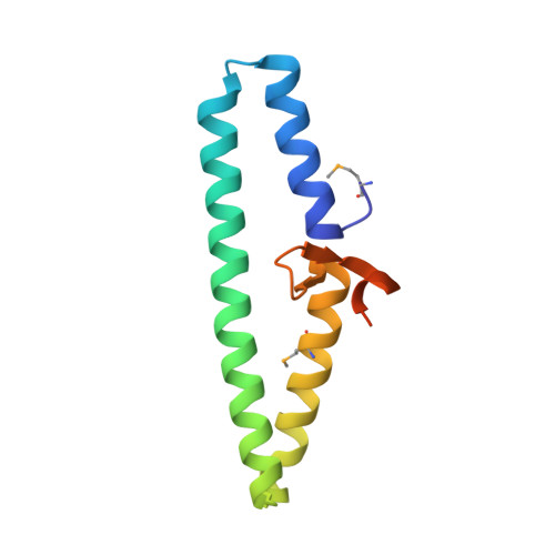
    CRISPR (clustered regularly interspaced short palindromic repeat)-Cas adaptive immune systems are prevalent defense mechanisms in bacteria and archaea. They provide sequence-specific detection and neutralization of foreign nucleic acids such as bacteriophages and plasmids. One mechanism by which phages and other mobile genetic elements are able to overcome the CRISPR-Cas system is through the expression of anti-CRISPR proteins. Over 20 different families of anti-CRISPR proteins have been described, each of which inhibits a particular type of CRISPR-Cas system. In this work, we determined the structure of type I-E anti-CRISPR protein AcrE1 by X-ray crystallography. We show that AcrE1 binds to the CRISPR-associated helicase/nuclease Cas3 and that the C-terminal region of the anti-CRISPR protein is important for its inhibitory activity. We further show that AcrE1 can convert the endogenous type I-E CRISPR system into a programmable transcriptional repressor. IMPORTANCE The CRISPR-Cas immune system provides bacteria with resistance to invasion by potentially harmful viruses, plasmids, and other foreign mobile genetic elements. This study presents the first structural and mechanistic insight into a phage-encoded protein that inactivates the type I-E CRISPR-Cas system in Pseudomonas aeruginosa The interaction of this anti-CRISPR protein with the CRISPR-associated helicase/nuclease proteins Cas3 shuts down the CRISPR-Cas system and protects phages carrying this gene from destruction. This interaction also allows the repurposing of the endogenous type I-E CRISPR system into a programmable transcriptional repressor, providing a new biotechnological tool for genetic studies of bacteria encoding this type I-E CRISPR-Cas system.


  • Organizational Affiliation
    • Department of Biochemistry, University of Toronto, Toronto, Ontario, Canada.

Macromolecule Content 

  • Total Structure Weight: 38.15 kDa 
  • Atom Count: 2,358 
  • Modeled Residue Count: 282 
  • Deposited Residue Count: 324 
  • Unique protein chains: 1

Macromolecules

Find similar proteins by:|  3D Structure
Entity ID: 1
MoleculeChains  Sequence LengthOrganismDetailsImage
Uncharacterized protein
A, B, C
108Pseudomonas phage JBD5Mutation(s): 0 
Gene Names: JBD5_034
UniProt
Find proteins for L7P7L6 (Pseudomonas phage JBD5)
Explore L7P7L6 
Go to UniProtKB:  L7P7L6
Entity Groups
Sequence Clusters30% Identity50% Identity70% Identity90% Identity95% Identity100% Identity
UniProt GroupL7P7L6
Sequence Annotations
Expand
Reference Sequence

Small Molecules

Ligands 3 Unique
IDChains Name / Formula / InChI Key2D Diagram3D Interactions
PG4

Query on PG4


Download:Ideal Coordinates CCD File
F [auth A]TETRAETHYLENE GLYCOL
C8 H18 O5
UWHCKJMYHZGTIT-UHFFFAOYSA-N
GOL

Query on GOL


Download:Ideal Coordinates CCD File
D [auth A],
E [auth A]
GLYCEROL
C3 H8 O3
PEDCQBHIVMGVHV-UHFFFAOYSA-N
BR

Query on BR


Download:Ideal Coordinates CCD File
G [auth B],
H [auth B],
I [auth B],
J [auth B],
K [auth C]
BROMIDE ION
Br
CPELXLSAUQHCOX-UHFFFAOYSA-M
Modified Residues  1 Unique
IDChains TypeFormula2D DiagramParent
MSE
Query on MSE
A, B, C
L-PEPTIDE LINKINGC5 H11 N O2 SeMET

Experimental Data & Validation

Experimental Data

  • Method: X-RAY DIFFRACTION
  • Resolution: 2.50 Å
  • R-Value Free:  0.221 (Depositor), 0.226 (DCC) 
  • R-Value Work:  0.179 (Depositor), 0.189 (DCC) 
  • R-Value Observed: 0.181 (Depositor) 
Space Group: C 1 2 1
Unit Cell:
Length ( Å )Angle ( ˚ )
a = 98.137α = 90
b = 63.959β = 100.94
c = 59.701γ = 90
Software Package:
Software NamePurpose
PHENIXrefinement
PDB_EXTRACTdata extraction
XDSdata reduction
XDSdata scaling
PHENIXphasing

Structure Validation

View Full Validation Report


Entry History 

& Funding Information

Deposition Data


Funding OrganizationLocationGrant Number
Canada--

Revision History  (Full details and data files)

  • Version 1.0: 2018-08-29
    Type: Initial release
  • Version 1.1: 2019-03-20
    Changes: Data collection, Database references
  • Version 1.2: 2024-11-20
    Changes: Data collection, Database references, Refinement description, Structure summary