Defining the Specificity of Carbohydrate-Protein Interactions by Quantifying Functional Group Contributions.
Sood, A., Gerlits, O.O., Ji, Y., Bovin, N.V., Coates, L., Woods, R.J.(2018) J Chem Inf Model 58: 1889-1901
- PubMed: 30086239 Search on PubMedSearch on PubMed Central
- DOI: https://doi.org/10.1021/acs.jcim.8b00120
- Primary Citation Related Structures:
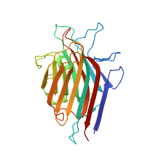
6AQ6 - PubMed Abstract:
Protein-carbohydrate interactions are significant in a wide range of biological processes, disruption of which has been implicated in many different diseases. The capability of glycan-binding proteins (GBPs) to specifically bind to the corresponding glycans allows GBPs to be utilized in glycan biomarker detection or conversely to serve as targets for therapeutic intervention. However, understanding the structural origins of GBP specificity has proven to be challenging due to their typically low binding affinities (mM) and their potential to display broad or complex specificities. Here we perform molecular dynamics (MD) simulations and post-MD energy analyses with the Poisson-Boltzmann and generalized Born solvent models (MM-PB/GBSA) of the Erythrina cristagalli lectin (ECL) with its known ligands, and with new cocrystal structures reported herein. While each MM-PB/GBSA parametrization resulted in different estimates of the desolvation free energy, general trends emerged that permit us to define GBP binding preferences in terms of ligand substructure specificity. Additionally, we have further decomposed the theoretical interaction energies into contributions made between chemically relevant functional groups. Based on these contributions, the functional groups in each ligand can be assembled into a pharmacophore comprised of groups that are either critical for binding, or enhance binding, or are noninteracting. It is revealed that the pharmacophore for ECL consists of the galactopyranose (Gal) ring atoms along with C6 and the O3 and O4 hydroxyl groups. This approach provides a convenient method for identifying and quantifying the glycan pharmacophore and provides a novel method for interpreting glycan specificity that is independent of residue-level glycan nomenclature. A pharmacophore approach to defining specificity is readily transferable to molecular design software and, therefore, may be particularly useful in designing therapeutics (glycomimetics) that target GBPs.
- Complex Carbohydrate Research Center , University of Georgia , Athens , Georgia 30602 , United States.
Organizational Affiliation: