6AO4

Crystal structure of the human gasdermin D C-terminal domain


Experimental Data Snapshot

  • Method: X-RAY DIFFRACTION
  • Resolution: 2.90 Å
  • R-Value Free: 0.290 
  • R-Value Work: 0.232 
  • R-Value Observed: 0.236 

wwPDB Validation   3D Report Full Report


This is version 1.3 of the entry. See complete history


Literature

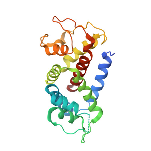
Structures of the Gasdermin D C-Terminal Domains Reveal Mechanisms of Autoinhibition.

Liu, Z.Wang, C.Rathkey, J.K.Yang, J.Dubyak, G.R.Abbott, D.W.Xiao, T.S.

(2018) Structure 26: 778-784.e3

  • DOI: https://doi.org/10.1016/j.str.2018.03.002
  • Primary Citation of Related Structures:  
    6AO3, 6AO4

  • PubMed Abstract: 

    Pyroptosis is an inflammatory form of programmed cell death that plays important roles in immune protection against infections and in inflammatory disorders. Gasdermin D (GSDMD) is an executor of pyroptosis upon cleavage by caspases-1/4/5/11 following canonical and noncanonical inflammasome activation. GSDMD N-terminal domain assembles membrane pores to induce cytolysis, whereas its C-terminal domain inhibits cell death through intramolecular association with the N domain. The molecular mechanisms of autoinhibition for GSDMD are poorly characterized. Here we report the crystal structures of the human and murine GSDMD C-terminal domains, which differ from those of the full-length murine GSDMA3 and the human GSDMB C-terminal domain. Mutations of GSDMD C-domain residues predicted to locate at its interface with the N-domain enhanced pyroptosis. Our results suggest that GSDMDs may employ a distinct mode of intramolecular domain interaction and autoinhibition, which may be relevant to its unique role in pyroptosis downstream of inflammasome activation.


  • Organizational Affiliation

    Department of Pathology, Case Western Reserve University, Cleveland, OH 44106, USA.


Macromolecules
Find similar proteins by:  (by identity cutoff)  |  3D Structure
Entity ID: 1
MoleculeChains Sequence LengthOrganismDetailsImage
Gasdermin-D209Homo sapiensMutation(s): 0 
Gene Names: GSDMDDFNA5LGSDMDC1FKSG10
UniProt & NIH Common Fund Data Resources
Find proteins for P57764 (Homo sapiens)
Explore P57764 
Go to UniProtKB:  P57764
PHAROS:  P57764
GTEx:  ENSG00000104518 
Entity Groups  
Sequence Clusters30% Identity50% Identity70% Identity90% Identity95% Identity100% Identity
UniProt GroupP57764
Sequence Annotations
Expand
  • Reference Sequence
Experimental Data & Validation

Experimental Data

  • Method: X-RAY DIFFRACTION
  • Resolution: 2.90 Å
  • R-Value Free: 0.290 
  • R-Value Work: 0.232 
  • R-Value Observed: 0.236 
  • Space Group: P 21 21 2
Unit Cell:
Length ( Å )Angle ( ˚ )
a = 47.75α = 90
b = 108.54β = 90
c = 45.91γ = 90
Software Package:
Software NamePurpose
PHENIXrefinement
XDSdata reduction
XSCALEdata scaling
PHASERphasing

Structure Validation

View Full Validation Report


Entry History & Funding Information

Deposition Data


Funding OrganizationLocationGrant Number
National Institutes of Health/National Institute of Arthritis and Musculoskeletal and Skin Diseases (NIH/NIAMS)United StatesAR069908

Revision History  (Full details and data files)

  • Version 1.0: 2018-04-11
    Type: Initial release
  • Version 1.1: 2018-05-09
    Changes: Data collection, Database references
  • Version 1.2: 2019-12-11
    Changes: Author supporting evidence
  • Version 1.3: 2023-10-04
    Changes: Data collection, Database references, Refinement description