6AMQ | pdb_00006amq

Crystal structure of the DNA polymerase III subunit beta from Enterobacter cloacae


Experimental Data Snapshot

  • Method: X-RAY DIFFRACTION
  • Resolution: 2.67 Å
  • R-Value Free: 
    0.300 (Depositor), 0.301 (DCC) 
  • R-Value Work: 
    0.246 (Depositor), 0.251 (DCC) 
  • R-Value Observed: 
    0.249 (Depositor) 

Starting Model: experimental
View more details

wwPDB Validation 3D Report Full Report

Validation slider image for 6AMQ

This is version 1.5 of the entry. See complete history

Literature

Crystal structures and biochemical characterization of DNA sliding clamps from three Gram-negative bacterial pathogens.

McGrath, A.E.Martyn, A.P.Whittell, L.R.Dawes, F.E.Beck, J.L.Dixon, N.E.Kelso, M.J.Oakley, A.J.

(2018) J Struct Biol 204: 396-405

  • DOI: https://doi.org/10.1016/j.jsb.2018.10.008
  • Primary Citation Related Structures: 
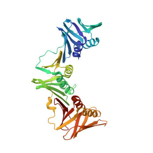
    6AMQ, 6AMS, 6AP4

  • PubMed Abstract: 

    Bacterial sliding clamps bind to DNA and act as protein-protein interaction hubs for several proteins involved in DNA replication and repair. The partner proteins all bind to a common pocket on sliding clamps via conserved linear peptide sequence motifs, which suggest the pocket as an attractive target for development of new antibiotics. Herein we report the X-ray crystal structures and biochemical characterization of β sliding clamps from the Gram-negative pathogens Pseudomonas aeruginosa, Acinetobacter baumannii and Enterobacter cloacae. The structures reveal close similarity between the pathogen and Escherichia coli clamps and similar patterns of binding to linear clamp-binding motif peptides. The results suggest that linear motif-sliding clamp interactions are well conserved and an antibiotic targeting the sliding clamp should have broad-spectrum activity against Gram-negative pathogens.


  • Organizational Affiliation
    • Molecular Horizons and School of Chemistry and Molecular Bioscience, University of Wollongong, and Illawarra Health and Medical Research Institute, Wollongong, New South Wales, Australia.

Macromolecule Content 

  • Total Structure Weight: 162.39 kDa 
  • Atom Count: 11,243 
  • Modeled Residue Count: 1,442 
  • Deposited Residue Count: 1,464 
  • Unique protein chains: 1

Macromolecules

Find similar proteins by:|  3D Structure
Entity ID: 1
MoleculeChains  Sequence LengthOrganismDetailsImage
DNA polymerase III subunit beta
A, B, C, D
366Enterobacter cloacae EcWSU1Mutation(s): 0 
Gene Names: dnaNEcWSU1_00002
UniProt
Find proteins for G8LES0 (Enterobacter ludwigii)
Explore G8LES0 
Go to UniProtKB:  G8LES0
Entity Groups
Sequence Clusters30% Identity50% Identity70% Identity90% Identity95% Identity100% Identity
UniProt GroupG8LES0
Sequence Annotations
Expand
Reference Sequence

Experimental Data & Validation

Experimental Data

  • Method: X-RAY DIFFRACTION
  • Resolution: 2.67 Å
  • R-Value Free:  0.300 (Depositor), 0.301 (DCC) 
  • R-Value Work:  0.246 (Depositor), 0.251 (DCC) 
  • R-Value Observed: 0.249 (Depositor) 
Space Group: P 1
Unit Cell:
Length ( Å )Angle ( ˚ )
a = 66.32α = 110.98
b = 79.229β = 91.64
c = 81.218γ = 89.96
Software Package:
Software NamePurpose
REFMACrefinement
SCALEPACKdata scaling
PDB_EXTRACTdata extraction
DENZOdata reduction
PHASERphasing

Structure Validation

View Full Validation Report


Entry History 

& Funding Information

Deposition Data


Funding OrganizationLocationGrant Number
Australian Research Council (ARC)AustraliaDP110100660
National Health and Medical Research Council (NHMRC, Australia)AustraliaAPP1021479

Revision History  (Full details and data files)

  • Version 1.0: 2017-11-01
    Type: Initial release
  • Version 1.1: 2017-12-06
    Changes: Author supporting evidence
  • Version 1.2: 2018-11-07
    Changes: Data collection, Database references, Source and taxonomy
  • Version 1.3: 2018-12-26
    Changes: Data collection, Database references
  • Version 1.4: 2020-01-01
    Changes: Author supporting evidence
  • Version 1.5: 2023-10-04
    Changes: Data collection, Database references, Refinement description