6AMG | pdb_00006amg

cyt P460 of Nitrosomonas sp. AL212


Experimental Data Snapshot

  • Method: X-RAY DIFFRACTION
  • Resolution: 1.45 Å
  • R-Value Free: 
    0.158 (Depositor), 0.170 (DCC) 
  • R-Value Work: 
    0.141 (Depositor), 0.153 (DCC) 
  • R-Value Observed: 
    0.142 (Depositor) 

Starting Model: experimental
View more details

wwPDB Validation 3D Report Full Report

Validation slider image for 6AMG

Ligand Structure Quality Assessment 


This is version 2.2 of the entry. See complete history

Literature

The Eponymous Cofactors in Cytochrome P460s from Ammonia-Oxidizing Bacteria Are Iron Porphyrinoids Whose Macrocycles Are Dibasic.

Smith, M.A.Lancaster, K.M.

(2018) Biochemistry 57: 334-343

  • DOI: https://doi.org/10.1021/acs.biochem.7b00921
  • Primary Citation Related Structures: 
    6AMG

  • PubMed Abstract: 

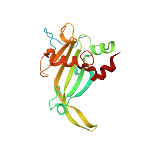
    The enzymes hydroxylamine oxidoreductase and cytochrome (cyt) P460 contain related unconventional "heme P460" cofactors. These cofactors are unusual in their inclusion of nonstandard cross-links between amino acid side chains and the heme macrocycle. Mutagenesis studies performed on the Nitrosomonas europaea cyt P460 that remove its lysine-heme cross-link show that the cross-link is key to defining the spectroscopic properties and kinetic competence of the enzyme. However, exactly how this cross-link confers these features remains unclear. Here we report the 1.45 Å crystal structure of cyt P460 from Nitrosomonas sp. AL212 and conclude that the cross-link does not lead to a change in hybridization of the heme carbon participating in the cross-link but rather enforces structural distortions to the macrocycle away from planarity. Time-dependent density functional theory coupled to experimental structural and spectroscopic analysis suggest that this geometric distortion is sufficient to define the spectroscopic properties of the heme P460 cofactor and provide clues toward establishing a relationship between heme P460 electronic structure and function.


  • Organizational Affiliation
    • Baker Laboratory, Department of Chemistry and Chemical Biology, Cornell University , Ithaca, New York 14853, United States.

Macromolecule Content 

  • Total Structure Weight: 44.64 kDa 
  • Atom Count: 2,893 
  • Modeled Residue Count: 318 
  • Deposited Residue Count: 392 
  • Unique protein chains: 1

Macromolecules

Find similar proteins by:|  3D Structure
Entity ID: 1
MoleculeChains  Sequence LengthOrganismDetailsImage
Cytochrome P460
A, B
196Nitrosomonas sp. AL212Mutation(s): 0 
Gene Names: NAL212_0896
UniProt
Find proteins for A0ACD6B8J9 (Nitrosomonas sp. AL212)
Explore A0ACD6B8J9 
Go to UniProtKB:  A0ACD6B8J9
Entity Groups
Sequence Clusters30% Identity50% Identity70% Identity90% Identity95% Identity100% Identity
UniProt GroupA0ACD6B8J9
Sequence Annotations
Expand
Reference Sequence

Experimental Data & Validation

Experimental Data

  • Method: X-RAY DIFFRACTION
  • Resolution: 1.45 Å
  • R-Value Free:  0.158 (Depositor), 0.170 (DCC) 
  • R-Value Work:  0.141 (Depositor), 0.153 (DCC) 
  • R-Value Observed: 0.142 (Depositor) 
Space Group: P 21 21 21
Unit Cell:
Length ( Å )Angle ( ˚ )
a = 47.267α = 90
b = 80.138β = 90
c = 108.962γ = 90
Software Package:
Software NamePurpose
PHENIXrefinement
XDSdata reduction
HKL-2000data scaling
PHENIXphasing

Structure Validation

View Full Validation Report


Ligand Structure Quality Assessment 


Entry History 

Deposition Data

Revision History  (Full details and data files)

  • Version 1.0: 2017-12-20
    Type: Initial release
  • Version 1.1: 2017-12-27
    Changes: Database references
  • Version 1.2: 2018-02-07
    Changes: Database references
  • Version 1.3: 2020-02-26
    Changes: Data collection
  • Version 2.0: 2021-03-10
    Changes: Atomic model, Data collection, Derived calculations, Non-polymer description, Structure summary
  • Version 2.1: 2023-10-04
    Changes: Data collection, Database references, Refinement description
  • Version 2.2: 2024-11-06
    Changes: Structure summary