Dimerization of MICU Proteins Controls Ca2+Influx through the Mitochondrial Ca2+Uniporter.
Xing, Y., Wang, M., Wang, J., Nie, Z., Wu, G., Yang, X., Shen, Y.(2019) Cell Rep 26: 1203-1212.e4
- PubMed: 30699349 Search on PubMed
- DOI: https://doi.org/10.1016/j.celrep.2019.01.022
- Primary Citation Related Structures:
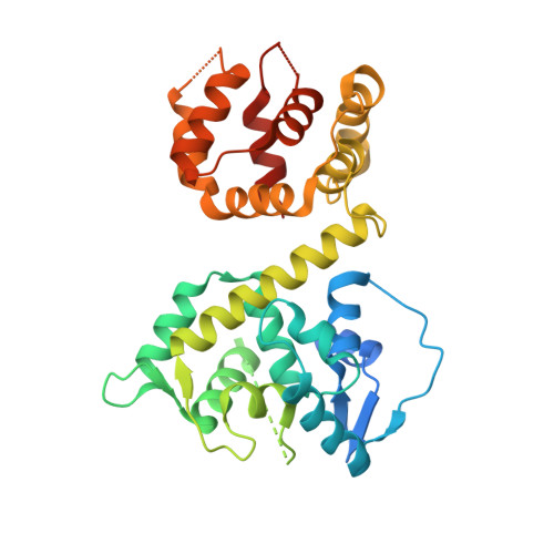
6AGH, 6AGI, 6AGJ - PubMed Abstract:
The mitochondrial Ca 2+ uniporter complex (MCUC) is responsible for Ca 2+ influx into the mitochondrial matrix, playing critical roles in various mitochondrial functions. Eukaryotic MCUC consists of multiple subunits, and its Ca 2+ influx activity is controlled by regulatory subunits, including mitochondrial Ca 2+ uptake 1 (MICU1) and its paralogs (MICU2 and MICU3). However, the underlying mechanism remains unclear. Here, we determined multiple crystal structures of MICU2 and MICU3 from Homo sapiens. Our data demonstrate that distinct MICU protein N-domains determine the specific type of MICU dimers that perform the opposing roles in mitochondrial Ca 2+ uptake at low cytosolic Ca 2+ levels. In contrast, at high cytosolic Ca 2+ levels, all MICU proteins undergo dimer rearrangement induced by Ca 2+ binding, which releases the suppression of the MCUC pore-forming subunit and promotes the influx of large amounts of Ca 2+ . Altogether, our results elucidate the delicate mechanism of mitochondrial Ca 2+ uptake regulation by MICU proteins.
- State Key Laboratory of Medicinal Chemical Biology and College of Life Sciences, Nankai University, 94 Weijin Road, Tianjin 300071, China.
Organizational Affiliation: