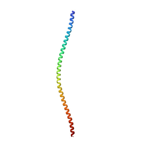
Crystal structure of the human glial fibrillary acidic protein 1B domain
Kim, B., Kim, S., Jin, M.S.(2018) Biochem Biophys Res Commun 503: 2899-2905
- PubMed: 30126635
- DOI: https://doi.org/10.1016/j.bbrc.2018.08.066
- Primary Citation of Related Structures:
6A9P - PubMed Abstract:
Glial fibrillary acidic protein (GFAP) is a homopolymeric type III intermediate filament (IF) that plays essential roles in cell migration, mitosis, development, and signaling in astrocytes and a specific type of glial cells. Its overexpression and genetic mutations lead to abnormal IF networks and accumulation of Rosenthal fibers, which results in the fatal neurodegenerative disorder Alexander disease. Herein, we present the first crystal structure of human GFAP spanning the central coiled-coil 1B domain at 2.5 Å resolution. The domain forms a tetramer comprising two equivalent parallel coiled-coil dimers that pack together in an antiparallel manner. Its assembly is stabilized by extensive networks of intermolecular hydrogen bonds, salt bridges, and hydrophobic interactions. Furthermore, mapping of the GFAP mutations associated with Alexander disease reveals that most involve residues buried in the core of the interface, and are likely to disrupt the intermolecular interactions and/or introduce steric clashes, thereby decreasing GFAP solubility and promoting aggregation. Based on our structural analysis and previous biochemical studies, we propose that GFAP assembles in the A11 mode in which coiled-coil 1B dimers lie in close axial proximity in an antiparallel fashion to provide a stable tetrameric platform for the organization of the GFAP filament.
- School of Life Sciences, GIST, 123 Cheomdan-gwagiro, Buk-gu, Gwangju 61005, Republic of Korea; Advanced Photonics Research Institute, GIST, 123 Cheomdan-gwagiro, Buk-gu, Gwangju 61005, Republic of Korea.
Organizational Affiliation:


