6A8U

PhoQ sensor domain (wild type): analysis of internal cavity


Experimental Data Snapshot

  • Method: X-RAY DIFFRACTION
  • Resolution: 1.85 Å
  • R-Value Free: 0.225 
  • R-Value Work: 0.169 
  • R-Value Observed: 0.172 

wwPDB Validation   3D Report Full Report


This is version 1.2 of the entry. See complete history


Literature

Identification of an internal cavity in the PhoQ sensor domain for PhoQ activity and SafA-mediated control.

Yoshitani, K.Ishii, E.Taniguchi, K.Sugimoto, H.Shiro, Y.Akiyama, Y.Kato, A.Utsumi, R.Eguchi, Y.

(2019) Biosci Biotechnol Biochem 83: 684-694

  • DOI: https://doi.org/10.1080/09168451.2018.1562879
  • Primary Citation of Related Structures:  
    6A8U, 6A8V

  • PubMed Abstract: 

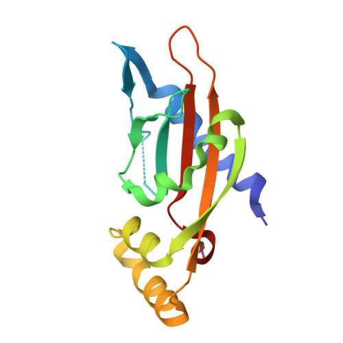
    The PhoQ/PhoP two-component signal transduction system is conserved in various Gram-negative bacteria and is often involved in the expression of virulence in pathogens. The small inner membrane protein SafA activates PhoQ in Escherichia coli independently from other known signals that control PhoQ activity. We have previously shown that SafA directly interacts with the sensor domain of the periplasmic region of PhoQ (PhoQ-SD) for activation, and that a D179R mutation in PhoQ-SD attenuates PhoQ activation by SafA. In this study, structural comparison of wild-type PhoQ-SD and D179R revealed a difference in the cavity (SD (sensory domain) pocket) found in the central core of this domain. This was the only structural difference between the two proteins. Site-directed mutagenesis of the residues surrounding the SD pocket has supported the SD pocket as a site involved in PhoQ activity. Furthermore, the SD pocket has also been shown to be involved in SafA-mediated PhoQ control.


  • Organizational Affiliation

    a Department of Bioscience , Graduate School of Agriculture, Kindai University , Nara , Japan.


Macromolecules
Find similar proteins by:  (by identity cutoff)  |  3D Structure
Entity ID: 1
MoleculeChains Sequence LengthOrganismDetailsImage
Sensor protein PhoQ
A, B
149Escherichia coli K-12Mutation(s): 0 
Gene Names: phoQb1129JW1115
EC: 2.7.13.3 (PDB Primary Data), 3.1.3 (PDB Primary Data)
UniProt
Find proteins for P23837 (Escherichia coli (strain K12))
Explore P23837 
Go to UniProtKB:  P23837
Entity Groups  
Sequence Clusters30% Identity50% Identity70% Identity90% Identity95% Identity100% Identity
UniProt GroupP23837
Sequence Annotations
Expand
  • Reference Sequence
Experimental Data & Validation

Experimental Data

  • Method: X-RAY DIFFRACTION
  • Resolution: 1.85 Å
  • R-Value Free: 0.225 
  • R-Value Work: 0.169 
  • R-Value Observed: 0.172 
  • Space Group: P 21 21 21
Unit Cell:
Length ( Å )Angle ( ˚ )
a = 38.03α = 90
b = 68.23β = 90
c = 112.896γ = 90
Software Package:
Software NamePurpose
PHENIXrefinement
HKL-2000data scaling
PDB_EXTRACTdata extraction
PHASERphasing
Cootmodel building

Structure Validation

View Full Validation Report


Entry History & Funding Information

Deposition Data


Funding OrganizationLocationGrant Number
Japan Society for the Promotion of ScienceJapanP20248012
Japan Society for the Promotion of ScienceJapanJP16K07681
Japan Society for the Promotion of ScienceJapanJP17J02501

Revision History  (Full details and data files)

  • Version 1.0: 2019-01-30
    Type: Initial release
  • Version 1.1: 2019-04-03
    Changes: Data collection, Database references
  • Version 1.2: 2023-11-22
    Changes: Data collection, Database references, Refinement description