6A8A

Ribokinase from Leishmania donovani with ATP


Experimental Data Snapshot

  • Method: X-RAY DIFFRACTION
  • Resolution: 1.80 Å
  • R-Value Free: 0.206 
  • R-Value Work: 0.182 
  • R-Value Observed: 0.183 

wwPDB Validation   3D Report Full Report


Ligand Structure Quality Assessment 


This is version 1.2 of the entry. See complete history


Literature

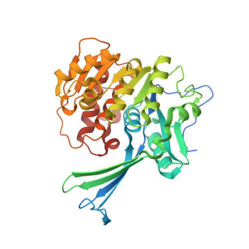
Unraveling structural insights of ribokinase from Leishmania donovani.

Gatreddi, S.Pillalamarri, V.Vasudevan, D.Addlagatta, A.Qureshi, I.A.

(2019) Int J Biol Macromol 136: 253-265

  • DOI: https://doi.org/10.1016/j.ijbiomac.2019.06.001
  • Primary Citation of Related Structures:  
    5ZWY, 6A8A, 6A8B, 6A8C

  • PubMed Abstract: 

    Ribokinase (RK) is an ATP dependent sugar kinase that enables the entry of ribose in the metabolism. Leishmania accumulates ribose into the cytosol through hydrolysis of nucleosides and by transport from the extracellular environment. Activation by RK is critical to mobilize the ribose into the metabolism of Leishmania. To understand the catalytic role, the crystal structure of RK (LdRK) from L. donovani was determined in the apo and complex forms with several nucleotides (ATP, AMPPCP and ADP) in the presence of Na + ion. The dual insertion of five amino acid stretches makes LdRK structurally unique from other reported structures of RKs. The structure of LdRK-ATP provided the basis for positioning of γ-phosphate of ATP by conserved -GAGD- motif. Liganded and unliganded structures of LdRK exists in similar conformation, which suggests binding of nucleotides does not make any significant conformational changes in nucleotide-bound structures. Substitution of a conserved asparagine with phenylalanine in ribose binding pocket differentiates the LdRK from other RKs. Glycerol molecule bound in the substrate binding pocket mimics the enzyme-substrate interactions but in turn, hampers the binding of ribose to LdRK. Comparative structural analysis revealed the flexibility of γ-phosphate, which adopts multiple conformations in the absence of divalent metal ion and ribose. Similar to other RKs, LdRK is also dependent on monovalent as well as divalent cations for its catalytic activity.


  • Organizational Affiliation

    Department of Biotechnology and Bioinformatics, School of Life Sciences, University of Hyderabad, Hyderabad 500 046, Telangana, India.


Macromolecules
Find similar proteins by:  (by identity cutoff)  |  3D Structure
Entity ID: 1
MoleculeChains Sequence LengthOrganismDetailsImage
Ribokinase
A, B
349Leishmania donovani BPK282A1Mutation(s): 0 
Gene Names: LDBPK_270430
EC: 2.7.1.15
UniProt
Find proteins for A0A3S7X0F5 (Leishmania donovani)
Explore A0A3S7X0F5 
Go to UniProtKB:  A0A3S7X0F5
Entity Groups  
Sequence Clusters30% Identity50% Identity70% Identity90% Identity95% Identity100% Identity
UniProt GroupA0A3S7X0F5
Sequence Annotations
Expand
  • Reference Sequence
Small Molecules
Ligands 4 Unique
IDChains Name / Formula / InChI Key2D Diagram3D Interactions
ATP
Query on ATP

Download Ideal Coordinates CCD File 
E [auth A],
M [auth B]
ADENOSINE-5'-TRIPHOSPHATE
C10 H16 N5 O13 P3
ZKHQWZAMYRWXGA-KQYNXXCUSA-N
GOL
Query on GOL

Download Ideal Coordinates CCD File 
C [auth A],
D [auth A]
GLYCEROL
C3 H8 O3
PEDCQBHIVMGVHV-UHFFFAOYSA-N
CL
Query on CL

Download Ideal Coordinates CCD File 
J [auth A]
K [auth A]
L [auth A]
Q [auth B]
R [auth B]
J [auth A],
K [auth A],
L [auth A],
Q [auth B],
R [auth B],
S [auth B],
T [auth B]
CHLORIDE ION
Cl
VEXZGXHMUGYJMC-UHFFFAOYSA-M
NA
Query on NA

Download Ideal Coordinates CCD File 
F [auth A]
G [auth A]
H [auth A]
I [auth A]
N [auth B]
F [auth A],
G [auth A],
H [auth A],
I [auth A],
N [auth B],
O [auth B],
P [auth B]
SODIUM ION
Na
FKNQFGJONOIPTF-UHFFFAOYSA-N
Experimental Data & Validation

Experimental Data

  • Method: X-RAY DIFFRACTION
  • Resolution: 1.80 Å
  • R-Value Free: 0.206 
  • R-Value Work: 0.182 
  • R-Value Observed: 0.183 
  • Space Group: P 61
Unit Cell:
Length ( Å )Angle ( ˚ )
a = 99.902α = 90
b = 99.902β = 90
c = 125.554γ = 120
Software Package:
Software NamePurpose
REFMACrefinement
XDSdata reduction
SCALAdata scaling
MOLREPphasing

Structure Validation

View Full Validation Report


Ligand Structure Quality Assessment 


Entry History 

Deposition Data

Revision History  (Full details and data files)

  • Version 1.0: 2019-07-10
    Type: Initial release
  • Version 1.1: 2019-09-11
    Changes: Data collection, Database references
  • Version 1.2: 2023-11-22
    Changes: Data collection, Database references, Derived calculations, Refinement description