Identification, functional characterization, and crystal structure determination of bacterial levoglucosan dehydrogenase.
Sugiura, M., Nakahara, M., Yamada, C., Arakawa, T., Kitaoka, M., Fushinobu, S.(2018) J Biological Chem 293: 17375-17386
- PubMed: 30224354 Search on PubMedSearch on PubMed Central
- DOI: https://doi.org/10.1074/jbc.RA118.004963
- Primary Citation Related Structures:
6A3F, 6A3G, 6A3I, 6A3J - PubMed Abstract:
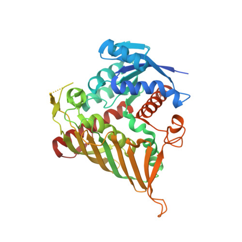
Levoglucosan is the 1,6-anhydrosugar of d-glucose formed by pyrolysis of glucans and is found in the environment and industrial waste. Two types of microbial levoglucosan metabolic pathways are known. Although the eukaryotic pathway involving levoglucosan kinase has been well-studied, the bacterial pathway involving levoglucosan dehydrogenase (LGDH) has not been well-investigated. Here, we identified and cloned the lgdh gene from the bacterium Pseudarthrobacter phenanthrenivorans and characterized the recombinant protein. The enzyme exhibited high substrate specificity toward levoglucosan and NAD + for the oxidative reaction and was confirmed to be LGDH. LGDH also showed weak activities (∼4%) toward l-sorbose and 1,5-anhydro-d-glucitol. The reverse (reductive) reaction using 3-keto-levoglucosan and NADH exhibited significantly lower K m and higher k cat values than those of the forward reaction. The crystal structures of LGDH in the apo and complex forms with NADH, NADH + levoglucosan, and NADH + l-sorbose revealed that LGDH has a typical fold of Gfo/Idh/MocA family proteins, similar to those of scyllo -inositol dehydrogenase, aldose-aldose oxidoreductase, 1,5-anhydro-d-fructose reductase, and glucose-fructose oxidoreductase. The crystal structures also disclosed that the active site of LGDH is distinct from those of these enzymes. The LGDH active site extensively recognized the levoglucosan molecule with six hydrogen bonds, and the C3 atom of levoglucosan was closely located to the C4 atom of NADH nicotinamide. Our study is the first molecular characterization of LGDH, providing evidence for C3-specific oxidation and representing a starting point for future biotechnological use of LGDH and levoglucosan-metabolizing bacteria.
- From the Department of Biotechnology and.
Organizational Affiliation: