6A2W | pdb_00006a2w

Crystal structure of fucoxanthin chlorophyll a/c complex from Phaeodactylum tricornutum


Experimental Data Snapshot

  • Method: X-RAY DIFFRACTION
  • Resolution: 1.80 Å
  • R-Value Free: 
    0.197 (Depositor), 0.195 (DCC) 
  • R-Value Work: 
    0.175 (Depositor), 0.173 (DCC) 

wwPDB Validation 3D Report Full Report

Validation slider image for 6A2W

Ligand Structure Quality Assessment 


This is version 3.0 of the entry. See complete history

Literature

Structural basis for blue-green light harvesting and energy dissipation in diatoms.

Wang, W.Yu, L.J.Xu, C.Tomizaki, T.Zhao, S.Umena, Y.Chen, X.Qin, X.Xin, Y.Suga, M.Han, G.Kuang, T.Shen, J.R.

(2019) Science 363

  • DOI: https://doi.org/10.1126/science.aav0365
  • Primary Citation Related Structures: 
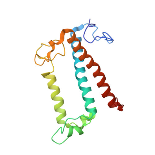
    6A2W

  • PubMed Abstract: 

    Diatoms are abundant photosynthetic organisms in aquatic environments and contribute 40% of its primary productivity. An important factor that contributes to the success of diatoms is their fucoxanthin chlorophyll a/c-binding proteins (FCPs), which have exceptional light-harvesting and photoprotection capabilities. Here, we report the crystal structure of an FCP from the marine diatom Phaeodactylum tricornutum , which reveals the binding of seven chlorophylls (Chls) a, two Chls c, seven fucoxanthins (Fxs), and probably one diadinoxanthin within the protein scaffold. Efficient energy transfer pathways can be found between Chl a and c, and each Fx is surrounded by Chls, enabling the energy transfer and quenching via Fx highly efficient. The structure provides a basis for elucidating the mechanisms of blue-green light harvesting, energy transfer, and dissipation in diatoms.


  • Organizational Affiliation
    • Photosynthesis Research Center, Key Laboratory of Photobiology, Institute of Botany, Chinese Academy of Sciences, 100093 Beijing, China.

Macromolecule Content 

  • Total Structure Weight: 34.51 kDa 
  • Atom Count: 2,518 
  • Modeled Residue Count: 166 
  • Deposited Residue Count: 167 
  • Unique protein chains: 1

Macromolecules

Find similar proteins by:|  3D Structure
Entity ID: 1
MoleculeChains  Sequence LengthOrganismDetailsImage
Protein fucoxanthin chlorophyl a/c protein167Phaeodactylum tricornutum CCAP 1055/1Mutation(s): 0 
Membrane Entity: Yes 
UniProt
Find proteins for B7FRW2 (Phaeodactylum tricornutum (strain CCAP 1055/1))
Explore B7FRW2 
Go to UniProtKB:  B7FRW2
Entity Groups
Sequence Clusters30% Identity50% Identity70% Identity90% Identity95% Identity100% Identity
UniProt GroupB7FRW2
Sequence Annotations
Expand
Reference Sequence

Small Molecules

Ligands 11 Unique
IDChains Name / Formula / InChI Key2D Diagram3D Interactions
DGD

Query on DGD


Download:Ideal Coordinates CCD File
V [auth A]DIGALACTOSYL DIACYL GLYCEROL (DGDG)
C51 H96 O15
LDQFLSUQYHBXSX-HXXRYREZSA-N
CLA

Query on CLA


Download:Ideal Coordinates CCD File
L [auth A]
M [auth A]
O [auth A]
P [auth A]
Q [auth A]
L [auth A],
M [auth A],
O [auth A],
P [auth A],
Q [auth A],
R [auth A],
T [auth A]
CHLOROPHYLL A
C55 H72 Mg N4 O5
ATNHDLDRLWWWCB-AENOIHSZSA-M
LHG

Query on LHG


Download:Ideal Coordinates CCD File
W [auth A]1,2-DIPALMITOYL-PHOSPHATIDYL-GLYCEROLE
C38 H75 O10 P
BIABMEZBCHDPBV-MPQUPPDSSA-N
A86

Query on A86


Download:Ideal Coordinates CCD File
D [auth A]
E [auth A]
F [auth A]
G [auth A]
H [auth A]
D [auth A],
E [auth A],
F [auth A],
G [auth A],
H [auth A],
I [auth A],
J [auth A]
(3S,3'S,5R,5'R,6S,6'R,8'R)-3,5'-dihydroxy-8-oxo-6',7'-didehydro-5,5',6,6',7,8-hexahydro-5,6-epoxy-beta,beta-caroten-3'- yl acetate
C42 H58 O6
SJWWTRQNNRNTPU-XJUZQKKNSA-N
KC1

Query on KC1


Download:Ideal Coordinates CCD File
S [auth A]Chlorophyll c1
C35 H30 Mg N4 O5
DGNIJJSSARBJSH-QIEHNWLWSA-L
KC2

Query on KC2


Download:Ideal Coordinates CCD File
N [auth A]Chlorophyll c2
C35 H28 Mg N4 O5
QDRBYWCRXZZVLY-QIEHNWLWSA-L
DD6

Query on DD6


Download:Ideal Coordinates CCD File
K [auth A](3S,3'R,5R,6S,7cis)-7',8'-didehydro-5,6-dihydro-5,6-epoxy-beta,beta-carotene-3,3'-diol
C40 H54 O3
OGHZCSINIMWCSB-WMTIXGNLSA-N
LMT

Query on LMT


Download:Ideal Coordinates CCD File
U [auth A]DODECYL-BETA-D-MALTOSIDE
C24 H46 O11
NLEBIOOXCVAHBD-QKMCSOCLSA-N
SOG

Query on SOG


Download:Ideal Coordinates CCD File
AA [auth A],
X [auth A],
Y [auth A],
Z [auth A]
octyl 1-thio-beta-D-glucopyranoside
C14 H28 O5 S
CGVLVOOFCGWBCS-RGDJUOJXSA-N
CA

Query on CA


Download:Ideal Coordinates CCD File
B [auth A],
C [auth A]
CALCIUM ION
Ca
BHPQYMZQTOCNFJ-UHFFFAOYSA-N
UNL

Query on UNL


Download:Ideal Coordinates CCD File
BA [auth A]
CA [auth A]
DA [auth A]
EA [auth A]
FA [auth A]
BA [auth A],
CA [auth A],
DA [auth A],
EA [auth A],
FA [auth A],
GA [auth A],
HA [auth A]
Unknown ligand
BHPQYMZQTOCNFJ-UHFFFAOYSA-N

Experimental Data & Validation

Experimental Data

  • Method: X-RAY DIFFRACTION
  • Resolution: 1.80 Å
  • R-Value Free:  0.197 (Depositor), 0.195 (DCC) 
  • R-Value Work:  0.175 (Depositor), 0.173 (DCC) 
Space Group: C 2 2 21
Unit Cell:
Length ( Å )Angle ( ˚ )
a = 47.749α = 90
b = 115.718β = 90
c = 141.261γ = 90
Software Package:
Software NamePurpose
PHENIXrefinement
XDSdata reduction
XSCALEdata scaling
PDB_EXTRACTdata extraction
CRANK2phasing

Structure Validation

View Full Validation Report


Ligand Structure Quality Assessment 


Entry History 

Deposition Data

Revision History  (Full details and data files)

  • Version 1.0: 2019-02-06
    Type: Initial release
  • Version 2.0: 2019-02-20
    Changes: Advisory, Atomic model, Data collection, Derived calculations
  • Version 2.1: 2019-02-27
    Changes: Data collection, Database references
  • Version 2.2: 2020-07-29
    Type: Remediation
    Reason: Carbohydrate remediation
    Changes: Derived calculations, Structure summary
  • Version 3.0: 2024-03-27
    Changes: Data collection, Database references, Non-polymer description, Structure summary