5ZWL

Crystal structure of the gamma - epsilon complex of photosynthetic cyanobacterial F1-ATPase


Experimental Data Snapshot

  • Method: X-RAY DIFFRACTION
  • Resolution: 1.98 Å
  • R-Value Free: 0.242 
  • R-Value Work: 0.205 
  • R-Value Observed: 0.207 

wwPDB Validation   3D Report Full Report


This is version 1.2 of the entry. See complete history


Literature

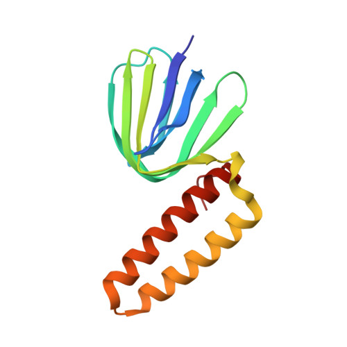
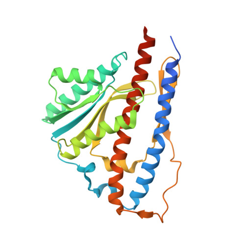
Structure of the gamma-epsilon complex of cyanobacterial F1-ATPase reveals a suppression mechanism of the gamma subunit on ATP hydrolysis in phototrophs.

Murakami, S.Kondo, K.Katayama, S.Hara, S.Sunamura, E.I.Yamashita, E.Groth, G.Hisabori, T.

(2018) Biochem J 475: 2925-2939

  • DOI: https://doi.org/10.1042/BCJ20180481
  • Primary Citation of Related Structures:  
    5ZWL

  • PubMed Abstract: 

    F 1 -ATPase forms the membrane-associated segment of F 0 F 1 -ATP synthase - the fundamental enzyme complex in cellular bioenergetics for ATP hydrolysis and synthesis. Here, we report a crystal structure of the central F 1 subcomplex, consisting of the rotary shaft γ subunit and the inhibitory ε subunit, from the photosynthetic cyanobacterium Thermosynechococcus elongatus BP-1, at 1.98 Å resolution. In contrast with their homologous bacterial and mitochondrial counterparts, the γ subunits of photosynthetic organisms harbour a unique insertion of 35-40 amino acids. Our structural data reveal that this region forms a β-hairpin structure along the central stalk. We identified numerous critical hydrogen bonds and electrostatic interactions between residues in the hairpin and the rest of the γ subunit. To elaborate the critical function of this β-hairpin in inhibiting ATP hydrolysis, the corresponding domain was deleted in the cyanobacterial F 1 subcomplex. Biochemical analyses of the corresponding α 3 β 3 γ complex confirm that the clinch of the hairpin structure plays a critical role and accounts for a significant interaction in the α 3 β 3 complex to induce ADP inhibition during ATP hydrolysis. In addition, we found that truncating the β-hairpin insertion structure resulted in a marked impairment of the interaction with the ε subunit, which binds to the opposite side of the γ subunit from the β-hairpin structure. Combined with structural analyses, our work provides experimental evidence supporting the molecular principle of how the insertion region of the γ subunit suppresses F 1 rotation during ATP hydrolysis.


  • Organizational Affiliation

    Department of Life Science, Tokyo Institute of Technology, Nagatsuta, Midori-ku, Yokohama 226-8501, Japan.


Macromolecules
Find similar proteins by:  (by identity cutoff)  |  3D Structure
Entity ID: 1
MoleculeChains Sequence LengthOrganismDetailsImage
ATP synthase epsilon chainA [auth E]138Thermosynechococcus vestitus BP-1Mutation(s): 0 
Gene Names: atpCatpEtlr0526
UniProt
Find proteins for Q8DLG7 (Thermosynechococcus vestitus (strain NIES-2133 / IAM M-273 / BP-1))
Explore Q8DLG7 
Go to UniProtKB:  Q8DLG7
Entity Groups  
Sequence Clusters30% Identity50% Identity70% Identity90% Identity95% Identity100% Identity
UniProt GroupQ8DLG7
Sequence Annotations
Expand
  • Reference Sequence
Find similar proteins by:  (by identity cutoff)  |  3D Structure
Entity ID: 2
MoleculeChains Sequence LengthOrganismDetailsImage
ATP synthase gamma chainB [auth G]280Thermosynechococcus vestitus BP-1Mutation(s): 2 
Gene Names: atpGatpCtll0385
UniProt
Find proteins for Q8DLU1 (Thermosynechococcus vestitus (strain NIES-2133 / IAM M-273 / BP-1))
Explore Q8DLU1 
Go to UniProtKB:  Q8DLU1
Entity Groups  
Sequence Clusters30% Identity50% Identity70% Identity90% Identity95% Identity100% Identity
UniProt GroupQ8DLU1
Sequence Annotations
Expand
  • Reference Sequence
Experimental Data & Validation

Experimental Data

  • Method: X-RAY DIFFRACTION
  • Resolution: 1.98 Å
  • R-Value Free: 0.242 
  • R-Value Work: 0.205 
  • R-Value Observed: 0.207 
  • Space Group: P 41 21 2
Unit Cell:
Length ( Å )Angle ( ˚ )
a = 71.556α = 90
b = 71.556β = 90
c = 204.969γ = 90
Software Package:
Software NamePurpose
PHENIXrefinement
HKL-2000data reduction
HKL-2000data scaling
SHELXDEphasing

Structure Validation

View Full Validation Report


Entry History & Funding Information

Deposition Data


Funding OrganizationLocationGrant Number
Japan Agency for Medical Research and Development (AMED)JapanJP18am0101072

Revision History  (Full details and data files)

  • Version 1.0: 2018-09-26
    Type: Initial release
  • Version 1.1: 2018-10-03
    Changes: Data collection, Database references
  • Version 1.2: 2024-03-27
    Changes: Data collection, Database references