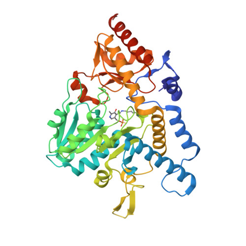
5ZS9

SufS from Bacillus subtilis in the resting state


Experimental Data Snapshot

  • Method: X-RAY DIFFRACTION
  • Resolution: 2.80 Å
  • R-Value Free: 0.237 
  • R-Value Work: 0.180 
  • R-Value Observed: 0.183 

wwPDB Validation   3D Report Full Report


This is version 1.3 of the entry. See complete history


Literature

Snapshots of PLP-substrate and PLP-product external aldimines as intermediates in two types of cysteine desulfurase enzymes.

Nakamura, R.Hikita, M.Ogawa, S.Takahashi, Y.Fujishiro, T.

(2020) FEBS J 287: 1138-1154

  • DOI: https://doi.org/10.1111/febs.15081
  • Primary Citation of Related Structures:  
    5WT2, 5WT4, 5ZS9, 5ZSK, 5ZSO, 5ZSP, 5ZST, 6KFZ, 6KG0, 6KG1

  • PubMed Abstract: 
  • Cysteine desulfurase enzymes catalyze sulfur mobilization from l-cysteine to sulfur-containing biomolecules such as iron-sulfur (Fe-S) clusters and thio-tRNAs. The enzymes utilize the cofactor pyridoxal-5'-phosphate (PLP), which forms the external substrate- and product-aldimines and ketimines during catalysis and are grouped into two types (I and II) based on their different catalytic loops ...

    Cysteine desulfurase enzymes catalyze sulfur mobilization from l-cysteine to sulfur-containing biomolecules such as iron-sulfur (Fe-S) clusters and thio-tRNAs. The enzymes utilize the cofactor pyridoxal-5'-phosphate (PLP), which forms the external substrate- and product-aldimines and ketimines during catalysis and are grouped into two types (I and II) based on their different catalytic loops. To clarify the structure-based catalytic mechanisms for each group, we determined the structures of the external substrate- and product-aldimines as catalytic intermediates of NifS (type I) and SufS (type II) that are involved in Fe-S cluster biosynthesis using X-ray crystallographic snapshot analysis. As a common intermediate structure, the thiol group of the PLP-l-cysteine external aldimine is stabilized by the conserved histidine adjacent to PLP through a polar interaction. This interaction makes the thiol group orientated for subsequent nucleophilic attack by a conserved cysteine residue on the catalytic loop in the state of PLP-l-cysteine ketimine, which is formed from the PLP-l-cysteine aldimine. Unlike the intermediates, structural changes of the loops were different between the type I and II enzymes. In the type I enzyme, conformational and topological change of the loop is necessary for nucleophilic attack by the cysteine. In contrast, the loop in type II cysteine desulfurase enzymes showed no large conformational change; rather, it might possibly orient the thiol group of the catalytic cysteine for nucleophilic attack toward PLP-l-cysteine. The present structures allow a revision of the catalytic mechanism and may provide a clue for consideration of enzyme function, structural diversity, and evolution of cysteine desulfurase enzymes. DATABASE: Structural data are available in PDB database under the accession numbers 5WT2, 5WT4, 5ZSP, 5ZST, 5ZS9, 5ZSK, 5ZSO, 6KFZ, 6KG0, and 6KG1.


    Organizational Affiliation

    Department of Biochemistry and Molecular Biology, Graduate School of Science and Engineering, Saitama University, Saitama, Japan.


Macromolecules
Find similar proteins by:  (by identity cutoff)  |  3D Structure
Entity ID: 1
MoleculeChainsSequence LengthOrganismDetailsImage
Cysteine desulfurase SufS419Bacillus subtilis subsp. subtilis str. 168Mutation(s): 0 
Gene Names: sufScsdyurWBSU32690
EC: 2.8.1.7
UniProt
Find proteins for O32164 (Bacillus subtilis (strain 168))
Explore O32164 
Go to UniProtKB:  O32164
Entity Groups  
Sequence Clusters30% Identity50% Identity70% Identity90% Identity95% Identity100% Identity
UniProt GroupO32164
Protein Feature View
Expand
  • Reference Sequence
Small Molecules
Ligands 1 Unique
IDChainsName / Formula / InChI Key2D Diagram3D Interactions
PEG
Query on PEG

Download Ideal Coordinates CCD File 
B [auth A]DI(HYDROXYETHYL)ETHER
C4 H10 O3
MTHSVFCYNBDYFN-UHFFFAOYSA-N
 Ligand Interaction
Modified Residues  1 Unique
IDChainsTypeFormula2D DiagramParent
LLP
Query on LLP
A
L-PEPTIDE LINKINGC14 H22 N3 O7 PLYS
Experimental Data & Validation

Experimental Data

  • Method: X-RAY DIFFRACTION
  • Resolution: 2.80 Å
  • R-Value Free: 0.237 
  • R-Value Work: 0.180 
  • R-Value Observed: 0.183 
  • Space Group: P 31 2 1
Unit Cell:
Length ( Å )Angle ( ˚ )
a = 91.9α = 90
b = 91.9β = 90
c = 127.1γ = 120
Software Package:
Software NamePurpose
XSCALEdata scaling
PHENIXrefinement
PDB_EXTRACTdata extraction
PHASERphasing
XDSdata reduction

Structure Validation

View Full Validation Report


Entry History & Funding Information

Deposition Data


Funding OrganizationLocationGrant Number
Japan Society for the Promotion of ScienceJapan17K14510
Japan Society for the Promotion of ScienceJapan15H04472

Revision History  (Full details and data files)

  • Version 1.0: 2019-05-01
    Type: Initial release
  • Version 1.1: 2019-10-16
    Changes: Data collection, Database references
  • Version 1.2: 2019-10-23
    Changes: Data collection, Database references
  • Version 1.3: 2020-04-01
    Changes: Database references