


Co-translational folding of alpha-helical proteins: structural studies of intermediate-length variants of the lambda repressor
Hanazono, Y., Takeda, K., Miki, K.(2018) FEBS Open Bio 8: 1312-1321
Experimental Data Snapshot
Starting Model: experimental
View more details
wwPDB Validation 3D Report Full Report

(2018) FEBS Open Bio 8: 1312-1321
Entity ID: 1 | |||||
|---|---|---|---|---|---|
| Molecule | Chains | Sequence Length | Organism | Details | Image |
| Repressor protein cI,Maltose-binding periplasmic protein | 391 | Lambdavirus lambda, Escherichia coli K-12 This entity is chimeric | Mutation(s): 0 Gene Names: cI, lambdap88, malE, b4034, JW3994 | 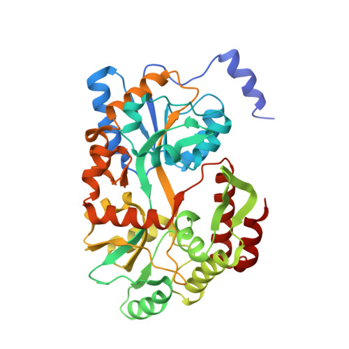 | |
UniProt | |||||
Entity Groups | |||||
| Sequence Clusters | 30% Identity50% Identity70% Identity90% Identity95% Identity100% Identity | ||||
| UniProt Groups | P03034P0AEX9 | ||||
Sequence AnnotationsExpand | |||||
Reference Sequence | |||||
| Ligands 1 Unique | |||||
|---|---|---|---|---|---|
| ID | Chains | Name / Formula / InChI Key | 2D Diagram | 3D Interactions | |
| CIT Download:Ideal Coordinates CCD File | C [auth A] | CITRIC ACID C6 H8 O7 KRKNYBCHXYNGOX-UHFFFAOYSA-N |  | ||
Entity ID: 2 | |||||
|---|---|---|---|---|---|
| ID | Chains | Name | Type/Class | 2D Diagram | 3D Interactions |
| PRD_900001 Query on PRD_900001 | B | alpha-maltose | Oligosaccharide / Nutrient |  |
| Length ( Å ) | Angle ( ˚ ) |
|---|---|
| a = 48.956 | α = 90 |
| b = 58.343 | β = 90 |
| c = 124.609 | γ = 90 |
| Software Name | Purpose |
|---|---|
| PHENIX | refinement |
| HKL-2000 | data reduction |
| HKL-2000 | data scaling |
| MOLREP | phasing |


