5ZBL

Crystal structure of type-I LOG from Corynebacterium glutamicum in complex with AMP


Experimental Data Snapshot

  • Method: X-RAY DIFFRACTION
  • Resolution: 2.30 Å
  • R-Value Free: 0.259 
  • R-Value Work: 0.196 
  • R-Value Observed: 0.199 

wwPDB Validation   3D Report Full Report


Ligand Structure Quality Assessment 


This is version 1.3 of the entry. See complete history


Literature

Structural insight into molecular mechanism of cytokinin activating protein from Pseudomonas aeruginosa PAO1.

Seo, H.Kim, K.J.

(2018) Environ Microbiol 20: 3214-3223

  • DOI: https://doi.org/10.1111/1462-2920.14287
  • Primary Citation of Related Structures:  
    5ZBJ, 5ZBK, 5ZBL

  • PubMed Abstract: 

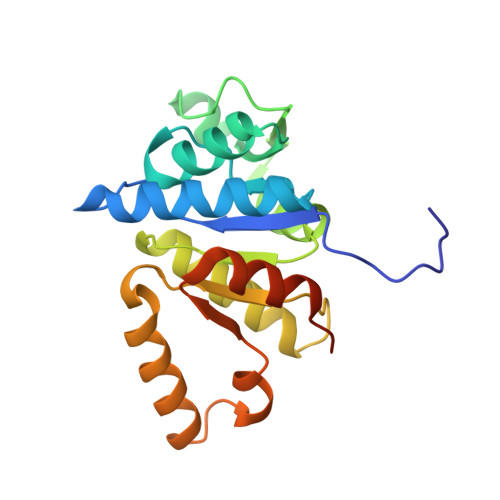
    Cytokinin (CK)-activating enzyme, called LOG, is a phosphoribohydrolase that hydrolyzes nucleotides into nucleobases and phosphoriboses. This reaction is a fascinating target for regulation of cellular active CK. However, misannotation of LOG as a lysine decarboxylase and the lack of detailed catalytic and substrate-binding mechanisms have prevented studies of LOG at a protein-level. In this study, we determined the crystal structure of PA4923 from Pseudomonas aeruginosa PAO1. The overall structure of PA4923 resembles those of type-I LOGs, and it exhibited phosphoribohydrolase activity against AMP. These observations indicated that PA4923 functions as an LOG. We also determined the PaLOG structure in complex with AMP and elucidated the detailed binding mode of LOG against the AMP substrate. Interestingly, PaLOG undergoes an open/closed conformational change upon binding AMP, during which the Glu74 residue located on the β3-β4 connecting loop flips 180° and moves 13 Å towards the AMP molecule. Structural and amino acid sequence comparisons of LOGs suggest that this conformational change upon substrate binding might be a common phenomenon in LOGs. In addition, based on our structural studies and the reported catalytic mechanism of nucleoside hydrolases, we proposed a catalytic mechanism for LOG in which an oxocarbenium ion-like transition state is formed.


  • Organizational Affiliation

    School of Life Sciences, KNU Creative BioResearch Group, Kyungpook National University, Daegu, 41566, Republic of Korea.


Macromolecules
Find similar proteins by:  (by identity cutoff)  |  3D Structure
Entity ID: 1
MoleculeChains Sequence LengthOrganismDetailsImage
Cytokinin riboside 5'-monophosphate phosphoribohydrolase
A, B, C, D
203Corynebacterium glutamicum ATCC 13032Mutation(s): 0 
Gene Names: Cg2612
EC: 3.2.2
UniProt
Find proteins for Q8NN34 (Corynebacterium glutamicum (strain ATCC 13032 / DSM 20300 / BCRC 11384 / JCM 1318 / LMG 3730 / NCIMB 10025))
Explore Q8NN34 
Go to UniProtKB:  Q8NN34
Entity Groups  
Sequence Clusters30% Identity50% Identity70% Identity90% Identity95% Identity100% Identity
UniProt GroupQ8NN34
Sequence Annotations
Expand
  • Reference Sequence
Small Molecules
Ligands 5 Unique
IDChains Name / Formula / InChI Key2D Diagram3D Interactions
AMP
Query on AMP

Download Ideal Coordinates CCD File 
P [auth C],
R [auth D]
ADENOSINE MONOPHOSPHATE
C10 H14 N5 O7 P
UDMBCSSLTHHNCD-KQYNXXCUSA-N
SO4
Query on SO4

Download Ideal Coordinates CCD File 
H [auth A],
N [auth B],
O [auth B],
T [auth D]
SULFATE ION
O4 S
QAOWNCQODCNURD-UHFFFAOYSA-L
PO4
Query on PO4

Download Ideal Coordinates CCD File 
G [auth A],
M [auth B]
PHOSPHATE ION
O4 P
NBIIXXVUZAFLBC-UHFFFAOYSA-K
GOL
Query on GOL

Download Ideal Coordinates CCD File 
F [auth A],
K [auth B],
L [auth B],
Q [auth C],
S [auth D]
GLYCEROL
C3 H8 O3
PEDCQBHIVMGVHV-UHFFFAOYSA-N
EDO
Query on EDO

Download Ideal Coordinates CCD File 
E [auth A],
I [auth B],
J [auth B]
1,2-ETHANEDIOL
C2 H6 O2
LYCAIKOWRPUZTN-UHFFFAOYSA-N
Experimental Data & Validation

Experimental Data

  • Method: X-RAY DIFFRACTION
  • Resolution: 2.30 Å
  • R-Value Free: 0.259 
  • R-Value Work: 0.196 
  • R-Value Observed: 0.199 
  • Space Group: P 21 21 21
Unit Cell:
Length ( Å )Angle ( ˚ )
a = 70.153α = 90
b = 70.714β = 90
c = 190.802γ = 90
Software Package:
Software NamePurpose
REFMACrefinement
PDB_EXTRACTdata extraction
HKL-2000data scaling
MOLREPphasing

Structure Validation

View Full Validation Report


Ligand Structure Quality Assessment 


Entry History 

Deposition Data

Revision History  (Full details and data files)

  • Version 1.0: 2018-06-27
    Type: Initial release
  • Version 1.1: 2018-10-17
    Changes: Data collection, Database references
  • Version 1.2: 2020-09-16
    Changes: Structure summary
  • Version 1.3: 2023-11-22
    Changes: Data collection, Database references, Refinement description