


Structural and biochemical analysis of a novel b-glucosidase EmGH1 from Erythrobacter marinus
Li, L., Zhao, Y., Hu, X.J., Li, J.X.To be published.
Experimental Data Snapshot
Starting Model: experimental
View more details
wwPDB Validation 3D Report Full Report

Entity ID: 1 | |||||
|---|---|---|---|---|---|
| Molecule | Chains | Sequence Length | Organism | Details | Image |
| EmGH1 | 785 | Aurantiacibacter marinus | Mutation(s): 0 Gene Names: AAV99_00160 | 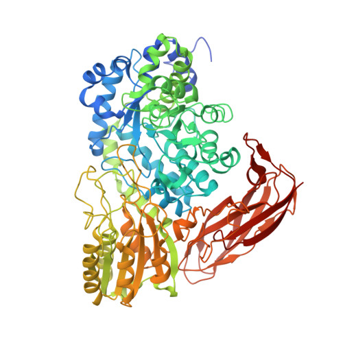 | |
UniProt | |||||
Find proteins for A0A0H0XV02 (Aurantiacibacter marinus) Explore A0A0H0XV02 Go to UniProtKB: A0A0H0XV02 | |||||
Entity Groups | |||||
| Sequence Clusters | 30% Identity50% Identity70% Identity90% Identity95% Identity100% Identity | ||||
| UniProt Group | A0A0H0XV02 | ||||
Sequence AnnotationsExpand | |||||
Reference Sequence | |||||
| Ligands 6 Unique | |||||
|---|---|---|---|---|---|
| ID | Chains | Name / Formula / InChI Key | 2D Diagram | 3D Interactions | |
| DTT Download:Ideal Coordinates CCD File | C [auth A], R [auth B] | 2,3-DIHYDROXY-1,4-DITHIOBUTANE C4 H10 O2 S2 VHJLVAABSRFDPM-IMJSIDKUSA-N |  | ||
| BEN Download:Ideal Coordinates CCD File | S [auth B] | BENZAMIDINE C7 H8 N2 PXXJHWLDUBFPOL-UHFFFAOYSA-N |  | ||
| PEG Download:Ideal Coordinates CCD File | AA [auth B] BA [auth B] N [auth A] O [auth A] P [auth A] | DI(HYDROXYETHYL)ETHER C4 H10 O3 MTHSVFCYNBDYFN-UHFFFAOYSA-N |  | ||
| GOL Download:Ideal Coordinates CCD File | G [auth A], H [auth A], Q [auth A], V [auth B], W [auth B] | GLYCEROL C3 H8 O3 PEDCQBHIVMGVHV-UHFFFAOYSA-N |  | ||
| EDO Download:Ideal Coordinates CCD File | I [auth A] J [auth A] K [auth A] L [auth A] M [auth A] | 1,2-ETHANEDIOL C2 H6 O2 LYCAIKOWRPUZTN-UHFFFAOYSA-N |  | ||
| MG Download:Ideal Coordinates CCD File | D [auth A], E [auth A], F [auth A], T [auth B], U [auth B] | MAGNESIUM ION Mg JLVVSXFLKOJNIY-UHFFFAOYSA-N |  | ||
| Length ( Å ) | Angle ( ˚ ) |
|---|---|
| a = 86.64 | α = 90 |
| b = 132.15 | β = 90 |
| c = 194.56 | γ = 90 |
| Software Name | Purpose |
|---|---|
| CrystalClear | data reduction |
| HKL-2000 | data scaling |
| PHASER | phasing |
| PHENIX | refinement |
| REFMAC | refinement |


