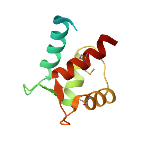
Crystal structure of the trimeric N-terminal domain of ciliate Euplotes octocarinatus centrin binding with calcium ions
Wang, W., Zhao, Y., Wang, H., Yang, B.(2018) Protein Sci 27: 1102-1108
- PubMed: 29607555
- DOI: https://doi.org/10.1002/pro.3418
- Primary Citation of Related Structures:
5Z1Q - PubMed Abstract:
Centrin is a member of the EF-hand superfamily of calcium-binding proteins, a highly conserved eukaryotic protein that binds to Ca 2+ . Its self-assembly plays a causative role in the fiber contraction that is associated with the cell division cycle and ciliogenesis. In this study, the crystal structure of N-terminal domain of ciliate Euplotes octocarinatus centrin (N-EoCen) was determined by using the selenomethionine single-wavelength anomalous dispersion method. The protein molecules formed homotrimers. Every protomer had two putative Ca 2+ ion-binding sites I and II, protomer A, and C bound one Ca 2+ ion, while protomer B bound two Ca 2+ ions. A novel binding site III was observed and the Ca 2+ ion was located at the center of the homotrimer. Several hydrogen bonds, electrostatic, and hydrophobic interactions between the protomers contributed to the formation of the oligomer. Structural studies provided insight into the foundation for centrin aggregation and the roles of calcium ions.
- Key Laboratory of Chemical Biology and Molecular Engineering of Education Ministry, Institute of Molecular Science, Shanxi University, Taiyuan, 030006, China.
Organizational Affiliation:


