5Z1Q | pdb_00005z1q

Crystal structures of the trimeric N-terminal domain of Ciliate Euplotes octocarinatus Centrin


Experimental Data Snapshot

  • Method: X-RAY DIFFRACTION
  • Resolution: 2.30 Å
  • R-Value Free: 
    0.224 (Depositor), 0.229 (DCC) 
  • R-Value Work: 
    0.184 (Depositor), 0.184 (DCC) 
  • R-Value Observed: 
    0.186 (Depositor) 

wwPDB Validation 3D Report Full Report

Validation slider image for 5Z1Q

This is version 1.2 of the entry. See complete history

Literature

Crystal structure of the trimeric N-terminal domain of ciliate Euplotes octocarinatus centrin binding with calcium ions

Wang, W.Zhao, Y.Wang, H.Yang, B.

(2018) Protein Sci 27: 1102-1108

  • DOI: https://doi.org/10.1002/pro.3418
  • Primary Citation Related Structures: 
    5Z1Q

  • PubMed Abstract: 

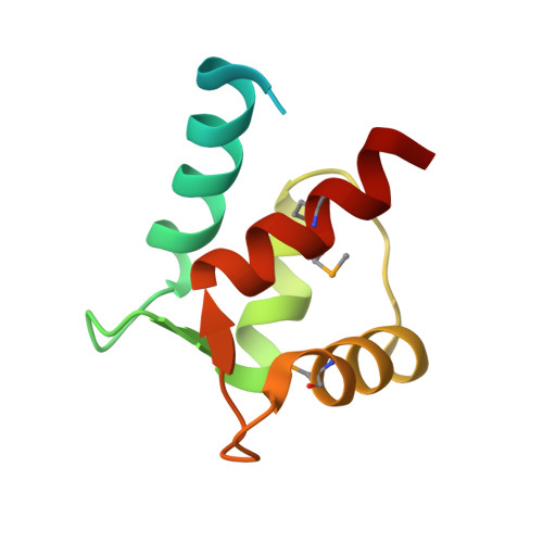
    Centrin is a member of the EF-hand superfamily of calcium-binding proteins, a highly conserved eukaryotic protein that binds to Ca 2+ . Its self-assembly plays a causative role in the fiber contraction that is associated with the cell division cycle and ciliogenesis. In this study, the crystal structure of N-terminal domain of ciliate Euplotes octocarinatus centrin (N-EoCen) was determined by using the selenomethionine single-wavelength anomalous dispersion method. The protein molecules formed homotrimers. Every protomer had two putative Ca 2+ ion-binding sites I and II, protomer A, and C bound one Ca 2+ ion, while protomer B bound two Ca 2+ ions. A novel binding site III was observed and the Ca 2+ ion was located at the center of the homotrimer. Several hydrogen bonds, electrostatic, and hydrophobic interactions between the protomers contributed to the formation of the oligomer. Structural studies provided insight into the foundation for centrin aggregation and the roles of calcium ions.


  • Organizational Affiliation
    • Key Laboratory of Chemical Biology and Molecular Engineering of Education Ministry, Institute of Molecular Science, Shanxi University, Taiyuan, 030006, China.

Macromolecule Content 

  • Total Structure Weight: 34.63 kDa 
  • Atom Count: 1,886 
  • Modeled Residue Count: 224 
  • Deposited Residue Count: 285 
  • Unique protein chains: 1

Macromolecules

Find similar proteins by:|  3D Structure
Entity ID: 1
MoleculeChains  Sequence LengthOrganismDetailsImage
Centrin protein
A, B, C
95Euplotes octocarinatusMutation(s): 0 
Gene Names: centrin
UniProt
Find proteins for Q9XZV2 (Euplotoides octocarinatus)
Explore Q9XZV2 
Go to UniProtKB:  Q9XZV2
Entity Groups
Sequence Clusters30% Identity50% Identity70% Identity90% Identity95% Identity100% Identity
UniProt GroupQ9XZV2
Sequence Annotations
Expand
Reference Sequence

Small Molecules

Modified Residues  1 Unique
IDChains TypeFormula2D DiagramParent
MSE
Query on MSE
A, B, C
L-PEPTIDE LINKINGC5 H11 N O2 SeMET

Experimental Data & Validation

Experimental Data

  • Method: X-RAY DIFFRACTION
  • Resolution: 2.30 Å
  • R-Value Free:  0.224 (Depositor), 0.229 (DCC) 
  • R-Value Work:  0.184 (Depositor), 0.184 (DCC) 
  • R-Value Observed: 0.186 (Depositor) 
Space Group: P 1
Unit Cell:
Length ( Å )Angle ( ˚ )
a = 42.945α = 106.88
b = 43.045β = 106.89
c = 43.087γ = 106.61
Software Package:
Software NamePurpose
PHENIXrefinement
HKL-3000data reduction
HKL-3000data scaling
PHENIXphasing

Structure Validation

View Full Validation Report


Entry History 

& Funding Information

Deposition Data


Funding OrganizationLocationGrant Number
National Natural Science Foundation of ChinaChina21601112
National Natural Science Foundation of ChinaChina21671125

Revision History  (Full details and data files)

  • Version 1.0: 2018-04-25
    Type: Initial release
  • Version 1.1: 2018-06-20
    Changes: Data collection, Database references
  • Version 1.2: 2024-11-20
    Changes: Data collection, Database references, Derived calculations, Structure summary