Positive Charges on the Surface of Thaumatin Are Crucial for the Multi-Point Interaction with the Sweet Receptor.
Masuda, T., Kigo, S., Mitsumoto, M., Ohta, K., Suzuki, M., Mikami, B., Kitabatake, N., Tani, F.(2018) Front Mol Biosci 5: 10-10
- PubMed: 29487853 Search on PubMedSearch on PubMed Central
- DOI: https://doi.org/10.3389/fmolb.2018.00010
- Primary Citation Related Structures:
5YYP, 5YYQ, 5YYR - PubMed Abstract:
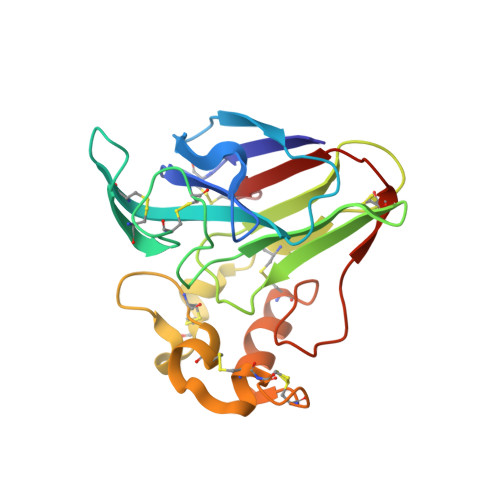
Thaumatin, an intensely sweet-tasting protein, elicits sweet taste with a threshold of only 50 nM. Previous studies from our laboratory suggested that the complex model between the T1R2-T1R3 sweet receptor and thaumatin depends critically on the complementarity of electrostatic potentials. In order to further validate this model, we focused on three lysine residues (Lys78, Lys106, and Lys137), which were expected to be part of the interaction sites. Three thaumatin mutants (K78A, K106A, and K137A) were prepared and their threshold values of sweetness were examined. The results showed that the sweetness of K106A was reduced by about three times and those of K78A and K137A were reduced by about five times when compared to wild-type thaumatin. The three-dimensional structures of these mutants were also determined by X-ray crystallographic analyses at atomic resolutions. The overall structures of mutant proteins were similar to that of wild-type but the electrostatic potentials around the mutated sites became more negative. Since the three lysine residues are located in 20-40 Å apart each other on the surface of thaumatin molecule, these results suggest the positive charges on the surface of thaumatin play a crucial role in the interaction with the sweet receptor, and are consistent with a large surface is required for interaction with the sweet receptor, as proposed by the multipoint interaction model named wedge model.
- Division of Food Science and Biotechnology, Graduate School of Agriculture, Kyoto University, Uji, Japan.
Organizational Affiliation: