5XYG

Structure of 6-aminohexanoate-oligomer hydrolase from Arthrobacter sp. KI72.


Experimental Data Snapshot

  • Method: X-RAY DIFFRACTION
  • Resolution: 1.60 Å
  • R-Value Free: 0.192 
  • R-Value Work: 0.161 
  • R-Value Observed: 0.162 

wwPDB Validation   3D Report Full Report


This is version 1.1 of the entry. See complete history


Literature

Structural basis of the correct subunit assembly, aggregation, and intracellular degradation of nylon hydrolase

Negoro, S.Shibata, N.Lee, Y.H.Takehara, I.Kinugasa, R.Nagai, K.Tanaka, Y.Kato, D.I.Takeo, M.Goto, Y.Higuchi, Y.

(2018) Sci Rep 8: 9725-9725

  • DOI: https://doi.org/10.1038/s41598-018-27860-w
  • Primary Citation of Related Structures:  
    5XYG, 5XYO, 5XYP, 5XYQ, 5XYS, 5XYT, 5Y0L, 5Y0M

  • PubMed Abstract: 

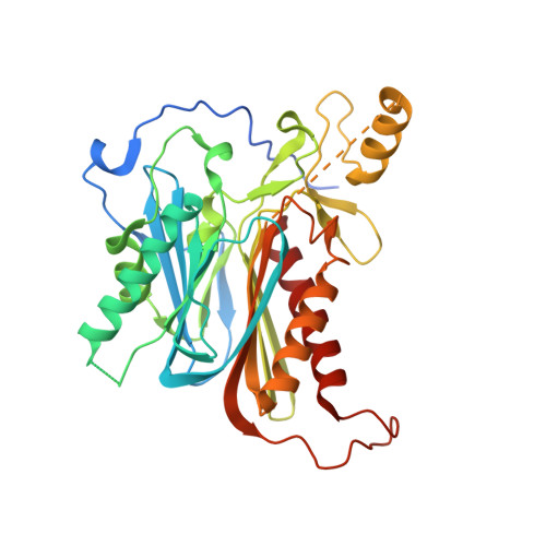
    Nylon hydrolase (NylC) is initially expressed as an inactive precursor (36 kDa). The precursor is cleaved autocatalytically at Asn266/Thr267 to generate an active enzyme composed of an α subunit (27 kDa) and a β subunit (9 kDa). Four αβ heterodimers (molecules A-D) form a doughnut-shaped quaternary structure. In this study, the thermostability of the parental NylC was altered by amino acid substitutions located at the A/D interface (D122G/H130Y/D36A/L137A) or the A/B interface (E263Q) and spanned a range of 47 °C. Considering structural, biophysical, and biochemical analyses, we discuss the structural basis of the stability of nylon hydrolase. From the analytical centrifugation data obtained regarding the various mutant enzymes, we conclude that the assembly of the monomeric units is dynamically altered by the mutations. Finally, we propose a model that can predict whether the fate of the nascent polypeptide will be correct subunit assembly, inappropriate protein-protein interactions causing aggregation, or intracellular degradation of the polypeptide.


  • Organizational Affiliation

    Department of Applied Chemistry, Graduate School of Engineering, University of Hyogo, Himeji, 671-2280, Japan. negoro@eng.u-hyogo.ac.jp.


Macromolecules
Find similar proteins by:  (by identity cutoff)  |  3D Structure
Entity ID: 1
MoleculeChains Sequence LengthOrganismDetailsImage
Endotype 6-aminohexanoat-oligomer hydrolase
A, B
355Arthrobacter sp.Mutation(s): 0 
UniProt
Find proteins for Q79F77 (Paenarthrobacter ureafaciens)
Explore Q79F77 
Go to UniProtKB:  Q79F77
Entity Groups  
Sequence Clusters30% Identity50% Identity70% Identity90% Identity95% Identity100% Identity
UniProt GroupQ79F77
Sequence Annotations
Expand
  • Reference Sequence
Small Molecules
Ligands 3 Unique
IDChains Name / Formula / InChI Key2D Diagram3D Interactions
SO4
Query on SO4

Download Ideal Coordinates CCD File 
E [auth A],
F [auth A],
I [auth B]
SULFATE ION
O4 S
QAOWNCQODCNURD-UHFFFAOYSA-L
GOL
Query on GOL

Download Ideal Coordinates CCD File 
C [auth A],
D [auth A]
GLYCEROL
C3 H8 O3
PEDCQBHIVMGVHV-UHFFFAOYSA-N
CL
Query on CL

Download Ideal Coordinates CCD File 
G [auth A],
H [auth A],
J [auth B],
K [auth B],
L [auth B]
CHLORIDE ION
Cl
VEXZGXHMUGYJMC-UHFFFAOYSA-M
Experimental Data & Validation

Experimental Data

  • Method: X-RAY DIFFRACTION
  • Resolution: 1.60 Å
  • R-Value Free: 0.192 
  • R-Value Work: 0.161 
  • R-Value Observed: 0.162 
  • Space Group: C 2 2 21
Unit Cell:
Length ( Å )Angle ( ˚ )
a = 70.844α = 90
b = 144.903β = 90
c = 129.045γ = 90
Software Package:
Software NamePurpose
REFMACrefinement
HKL-2000data reduction
HKL-2000data scaling
CNSphasing

Structure Validation

View Full Validation Report


Entry History & Funding Information

Deposition Data


Funding OrganizationLocationGrant Number
Japan Society for Promotion of ScienceJapan26289317

Revision History  (Full details and data files)

  • Version 1.0: 2018-11-21
    Type: Initial release
  • Version 1.1: 2023-11-22
    Changes: Data collection, Database references, Refinement description