5XWT | pdb_00005xwt

Crystal structure of PTPdelta Ig1-Fn1 in complex with SALM5 LRR-Ig


Experimental Data Snapshot

  • Method: X-RAY DIFFRACTION
  • Resolution: 4.18 Å
  • R-Value Free: 
    0.311 (Depositor), 0.318 (DCC) 
  • R-Value Work: 
    0.261 (Depositor), 0.274 (DCC) 
  • R-Value Observed: 
    0.263 (Depositor) 

wwPDB Validation 3D Report Full Report

Validation slider image for 5XWT

Ligand Structure Quality Assessment 


This is version 2.1 of the entry. See complete history

Literature

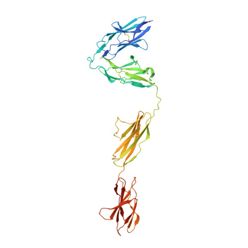
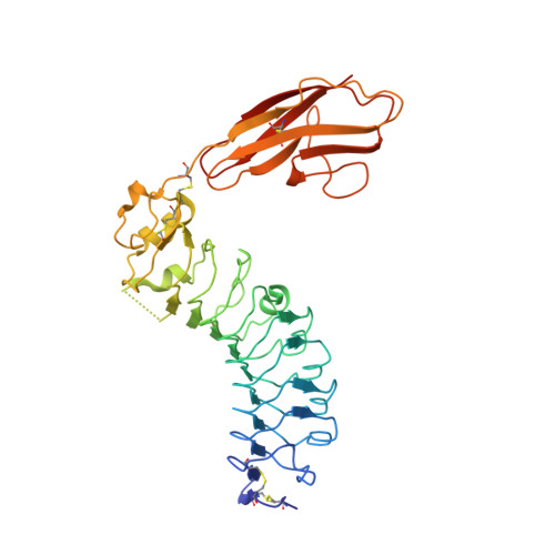
Structural basis of trans-synaptic interactions between PTP delta and SALMs for inducing synapse formation.

Goto-Ito, S.Yamagata, A.Sato, Y.Uemura, T.Shiroshima, T.Maeda, A.Imai, A.Mori, H.Yoshida, T.Fukai, S.

(2018) Nat Commun 9: 269-269

  • DOI: https://doi.org/10.1038/s41467-017-02417-z
  • Primary Citation Related Structures: 
    5XWS, 5XWT, 5XWU

  • PubMed Abstract: 

    Synapse formation is triggered by trans-synaptic interactions of cell adhesion molecules, termed synaptic organizers. Three members of type-II receptor protein tyrosine phosphatases (classified as type-IIa RPTPs; PTPδ, PTPσ and LAR) are known as presynaptic organizers. Synaptic adhesion-like molecules (SALMs) have recently emerged as a family of postsynaptic organizers. Although all five SALM isoforms can bind to the type-IIa RPTPs, only SALM3 and SALM5 reportedly have synaptogenic activities depending on their binding. Here, we report the crystal structures of apo-SALM5, and PTPδ-SALM2 and PTPδ-SALM5 complexes. The leucine-rich repeat (LRR) domains of SALMs interact with the second immunoglobulin-like (Ig) domain of PTPδ, whereas the Ig domains of SALMs interact with both the second and third Ig domains of PTPδ. Unexpectedly, the structures exhibit the LRR-mediated 2:2 complex. Our synaptogenic co-culture assay using site-directed SALM5 mutants demonstrates that presynaptic differentiation induced by PTPδ-SALM5 requires the dimeric property of SALM5.


  • Organizational Affiliation
    • Institute of Molecular and Cellular Biosciences, The University of Tokyo, Tokyo, 113-0032, Japan.

Macromolecule Content 

  • Total Structure Weight: 174.58 kDa 
  • Atom Count: 11,527 
  • Modeled Residue Count: 1,447 
  • Deposited Residue Count: 1,538 
  • Unique protein chains: 2

Macromolecules

Find similar proteins by:|  3D Structure
Entity ID: 1
MoleculeChains  Sequence LengthOrganismDetailsImage
Receptor-type tyrosine-protein phosphatase delta
A, C
402Mus musculusMutation(s): 0 
Gene Names: Ptprd
EC: 3.1.3.48
UniProt & NIH Common Fund Data Resources
Find proteins for Q64487 (Mus musculus)
Explore Q64487 
Go to UniProtKB:  Q64487
IMPC:  MGI:97812
Entity Groups
Sequence Clusters30% Identity50% Identity70% Identity90% Identity95% Identity100% Identity
UniProt GroupQ64487
Glycosylation
Glycosylation Sites: 2Go to GlyGen: Q64487-4
Sequence Annotations
Expand
Reference Sequence
Find similar proteins by:|  3D Structure
Entity ID: 2
MoleculeChains  Sequence LengthOrganismDetailsImage
Leucine-rich repeat and fibronectin type-III domain-containing protein 5
B, D
367Homo sapiensMutation(s): 0 
Gene Names: LRFN5C14orf146SALM5
UniProt & NIH Common Fund Data Resources
Find proteins for Q96NI6 (Homo sapiens)
Explore Q96NI6 
Go to UniProtKB:  Q96NI6
PHAROS:  Q96NI6
GTEx:  ENSG00000165379 
Entity Groups
Sequence Clusters30% Identity50% Identity70% Identity90% Identity95% Identity100% Identity
UniProt GroupQ96NI6
Glycosylation
Glycosylation Sites: 3Go to GlyGen: Q96NI6-1
Sequence Annotations
Expand
Reference Sequence

Oligosaccharides

Help  
Entity ID: 3
MoleculeChains Length2D Diagram GlycosylationD Interactions
2-acetamido-2-deoxy-beta-D-glucopyranose-(1-4)-2-acetamido-2-deoxy-beta-D-glucopyranose
E, F, H, I, J
2N-Glycosylation
Glycosylation Resources
GlyTouCan: G42666HT
GlyCosmos: G42666HT
GlyGen: G42666HT
Entity ID: 4
MoleculeChains Length2D Diagram GlycosylationD Interactions
2-acetamido-2-deoxy-beta-D-glucopyranose-(1-4)-2-acetamido-2-deoxy-beta-D-glucopyranose-(1-4)-2-acetamido-2-deoxy-beta-D-glucopyranose
G
3N-Glycosylation
Glycosylation Resources
GlyTouCan: G47362BJ
GlyCosmos: G47362BJ
GlyGen: G47362BJ

Biologically Interesting Molecules (External Reference) 

1 Unique

Experimental Data & Validation

Experimental Data

  • Method: X-RAY DIFFRACTION
  • Resolution: 4.18 Å
  • R-Value Free:  0.311 (Depositor), 0.318 (DCC) 
  • R-Value Work:  0.261 (Depositor), 0.274 (DCC) 
  • R-Value Observed: 0.263 (Depositor) 
Space Group: P 21 21 21
Unit Cell:
Length ( Å )Angle ( ˚ )
a = 98.26α = 90
b = 169.753β = 90
c = 210.946γ = 90
Software Package:
Software NamePurpose
PHENIXrefinement
HKL-2000data reduction
HKL-2000data scaling
PHASERphasing

Structure Validation

View Full Validation Report


Ligand Structure Quality Assessment 


Entry History 

Deposition Data

Revision History  (Full details and data files)

  • Version 1.0: 2018-06-06
    Type: Initial release
  • Version 2.0: 2020-07-29
    Type: Remediation
    Reason: Carbohydrate remediation
    Changes: Atomic model, Data collection, Derived calculations, Structure summary
  • Version 2.1: 2024-10-16
    Changes: Data collection, Database references, Structure summary