5XHG | pdb_00005xhg

Crystal structure of Trastuzumab Fab fragment bearing Ne-(o-azidobenzyloxycarbonyl)-L-lysine


Experimental Data Snapshot

  • Method: X-RAY DIFFRACTION
  • Resolution: 1.76 Å
  • R-Value Free: 
    0.199 (Depositor), 0.200 (DCC) 
  • R-Value Work: 
    0.173 (Depositor), 0.173 (DCC) 
  • R-Value Observed: 
    0.173 (Depositor) 

wwPDB Validation 3D Report Full Report

Validation slider image for 5XHG

Ligand Structure Quality Assessment 


This is version 1.3 of the entry. See complete history

Literature

Extensive Survey of Antibody Invariant Positions for Efficient Chemical Conjugation Using Expanded Genetic Codes.

Kato, A.Kuratani, M.Yanagisawa, T.Ohtake, K.Hayashi, A.Amano, Y.Kimura, K.Yokoyama, S.Sakamoto, K.Shiraishi, Y.

(2017) Bioconjug Chem 28: 2099-2108

  • DOI: https://doi.org/10.1021/acs.bioconjchem.7b00265
  • Primary Citation Related Structures: 
    5XHF, 5XHG

  • PubMed Abstract: 

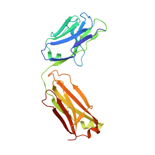
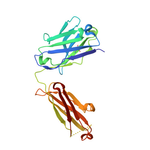
    The site-specific chemical conjugation of proteins, following synthesis with an expanded genetic code, promises to advance antibody-based technologies, including antibody drug conjugation and the creation of bispecific Fab dimers. The incorporation of non-natural amino acids into antibodies not only guarantees site specificity but also allows the use of bio-orthogonal chemistry. However, the efficiency of amino acid incorporation fluctuates significantly among different sites, thereby hampering the identification of useful conjugation sites. In this study, we applied the codon reassignment technology to achieve the robust and efficient synthesis of chemically functionalized antibodies containing N ε -(o-azidobenzyloxycarbonyl)-l-lysine (o-Az-Z-Lys) at defined positions. This lysine derivative has a bio-orthogonally reactive group at the end of a long side chain, enabling identification of multiple new positions in Fab-constant domains, allowing chemical conjugation with high efficiency. An X-ray crystallographic study of a Fab variant with o-Az-Z-Lys revealed high-level exposure of the azido group to solvent, with six of the identified positions subsequently used to engineer "Variabodies", a novel antibody format allowing various connections between two Fab molecules. Our findings indicated that some of the created Variabodies exhibited agonistic activity in cultured cells as opposed to the antagonistic nature of antibodies. These results showed that our approach greatly enhanced the availability of antibodies for chemical conjugation and might aid in the development of new therapeutic antibodies.


  • Organizational Affiliation
    • RIKEN Structural Biology Laboratory , 1-7-22 Suehiro-cho, Tsurumi-ku, Yokohama 230-0045, Japan.

Macromolecule Content 

  • Total Structure Weight: 95.04 kDa 
  • Atom Count: 7,919 
  • Modeled Residue Count: 861 
  • Deposited Residue Count: 868 
  • Unique protein chains: 2

Macromolecules

Find similar proteins by:|  3D Structure
Entity ID: 1
MoleculeChains  Sequence LengthOrganismDetailsImage
polypeptide(L)
A, C
214Mus musculusMutation(s): 0 
Find similar proteins by:|  3D Structure
Entity ID: 2
MoleculeChains  Sequence LengthOrganismDetailsImage
polypeptide(H)
B, D
220Mus musculusMutation(s): 0 

Small Molecules

Ligands 4 Unique
IDChains Name / Formula / InChI Key2D Diagram3D Interactions
OAZ

Query on OAZ


Download:Ideal Coordinates CCD File
E [auth A](2-azidophenyl)methyl hydrogen carbonate
C8 H7 N3 O2
YHJYQHHCLBKKKY-UHFFFAOYSA-N
PEG

Query on PEG


Download:Ideal Coordinates CCD File
I [auth B],
O [auth C]
DI(HYDROXYETHYL)ETHER
C4 H10 O3
MTHSVFCYNBDYFN-UHFFFAOYSA-N
SO4

Query on SO4


Download:Ideal Coordinates CCD File
F [auth A],
J [auth C],
P [auth D]
SULFATE ION
O4 S
QAOWNCQODCNURD-UHFFFAOYSA-L
EDO

Query on EDO


Download:Ideal Coordinates CCD File
G [auth A]
H [auth B]
K [auth C]
L [auth C]
M [auth C]
G [auth A],
H [auth B],
K [auth C],
L [auth C],
M [auth C],
N [auth C],
Q [auth D],
R [auth D],
S [auth D]
1,2-ETHANEDIOL
C2 H6 O2
LYCAIKOWRPUZTN-UHFFFAOYSA-N

Experimental Data & Validation

Experimental Data

  • Method: X-RAY DIFFRACTION
  • Resolution: 1.76 Å
  • R-Value Free:  0.199 (Depositor), 0.200 (DCC) 
  • R-Value Work:  0.173 (Depositor), 0.173 (DCC) 
  • R-Value Observed: 0.173 (Depositor) 
Space Group: P 1
Unit Cell:
Length ( Å )Angle ( ˚ )
a = 38.651α = 113.5
b = 79.891β = 92.82
c = 85.764γ = 102.84
Software Package:
Software NamePurpose
PHENIXrefinement
XDSdata reduction
XDSdata scaling
PHASERphasing

Structure Validation

View Full Validation Report


Ligand Structure Quality Assessment 


Entry History 

Deposition Data

Revision History  (Full details and data files)

  • Version 1.0: 2017-12-20
    Type: Initial release
  • Version 1.1: 2018-09-19
    Changes: Data collection, Derived calculations, Source and taxonomy, Structure summary
  • Version 1.2: 2019-12-25
    Changes: Source and taxonomy
  • Version 1.3: 2024-10-16
    Changes: Data collection, Database references, Derived calculations, Structure summary