


Crystal structure of peptidoglycan recognition protein (PGRP-S) at 2.45 A resolution
Shokeen, A., Sharma, P., Singh, P.K., Kaur, P., Sharma, S., Singh, T.P.To be published.
Experimental Data Snapshot
Starting Model: experimental
View more details
wwPDB Validation 3D Report Full Report

Entity ID: 1 | |||||
|---|---|---|---|---|---|
| Molecule | Chains | Sequence Length | Organism | Details | Image |
| Peptidoglycan recognition protein 1 | 171 | Camelus dromedarius | Mutation(s): 0 | 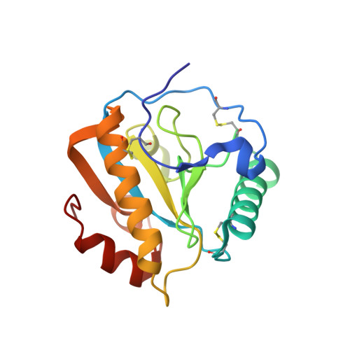 | |
UniProt | |||||
Entity Groups | |||||
| Sequence Clusters | 30% Identity50% Identity70% Identity90% Identity95% Identity100% Identity | ||||
| UniProt Group | Q9GK12 | ||||
Sequence AnnotationsExpand | |||||
Reference Sequence | |||||
| Ligands 2 Unique | |||||
|---|---|---|---|---|---|
| ID | Chains | Name / Formula / InChI Key | 2D Diagram | 3D Interactions | |
| TLA Download:Ideal Coordinates CCD File | F [auth C] | L(+)-TARTARIC ACID C4 H6 O6 FEWJPZIEWOKRBE-JCYAYHJZSA-N |  | ||
| GOL Download:Ideal Coordinates CCD File | E [auth C] | GLYCEROL C3 H8 O3 PEDCQBHIVMGVHV-UHFFFAOYSA-N |  | ||
| Length ( Å ) | Angle ( ˚ ) |
|---|---|
| a = 89.788 | α = 90 |
| b = 101.796 | β = 90 |
| c = 164.144 | γ = 90 |
| Software Name | Purpose |
|---|---|
| REFMAC | refinement |
| HKL-2000 | data reduction |
| SCALEPACK | data scaling |
| MOLREP | phasing |


