Computational design of a symmetrical beta-trefoil lectin with cancer cell binding activity.
Terada, D., Voet, A.R.D., Noguchi, H., Kamata, K., Ohki, M., Addy, C., Fujii, Y., Yamamoto, D., Ozeki, Y., Tame, J.R.H., Zhang, K.Y.J.(2017) Sci Rep 7: 5943-5943
- PubMed: 28724971 Search on PubMedSearch on PubMed Central
- DOI: https://doi.org/10.1038/s41598-017-06332-7
- Primary Citation Related Structures:
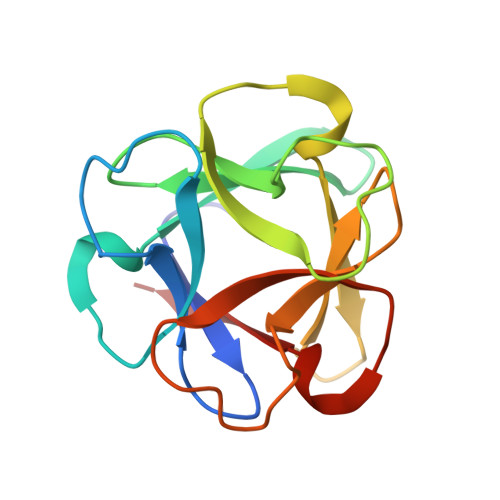
5XG5 - PubMed Abstract:
Computational protein design has advanced very rapidly over the last decade, but there remain few examples of artificial proteins with direct medical applications. This study describes a new artificial β-trefoil lectin that recognises Burkitt's lymphoma cells, and which was designed with the intention of finding a basis for novel cancer treatments or diagnostics. The new protein, called "Mitsuba", is based on the structure of the natural shellfish lectin MytiLec-1, a member of a small lectin family that uses unique sequence motifs to bind α-D-galactose. The three subdomains of MytiLec-1 each carry one galactose binding site, and the 149-residue protein forms a tight dimer in solution. Mitsuba (meaning "three-leaf" in Japanese) was created by symmetry constraining the structure of a MytiLec-1 subunit, resulting in a 150-residue sequence that contains three identical tandem repeats. Mitsuba-1 was expressed and crystallised to confirm the X-ray structure matches the predicted model. Mitsuba-1 recognises cancer cells that express globotriose (Galα(1,4)Galβ(1,4)Glc) on the surface, but the cytotoxicity is abolished.
- Graduate School of Medical Life Science, Yokohama City University, 1-7-29 Suehiro, Yokohama, Kanagawa, 230-0045, Japan.
Organizational Affiliation: