5XFU | pdb_00005xfu

Domain swapped dimer crystal structure of loop1 deletion mutant in Single-chain Monellin


Experimental Data Snapshot

  • Method: X-RAY DIFFRACTION
  • Resolution: 2.61 Å
  • R-Value Free: 
    0.294 (Depositor), 0.302 (DCC) 
  • R-Value Work: 
    0.260 (Depositor), 0.263 (DCC) 
  • R-Value Observed: 
    0.261 (Depositor) 

Starting Model: experimental
View more details

wwPDB Validation 3D Report Full Report

Validation slider image for 5XFU

This is version 1.2 of the entry. See complete history

Literature

Amino-acid composition after loop deletion drives domain swapping

Nandwani, N.Surana, P.Udgaonkar, J.B.Das, R.Gosavi, S.

(2017) Protein Sci 26: 1994-2002

  • DOI: https://doi.org/10.1002/pro.3237
  • Primary Citation Related Structures: 
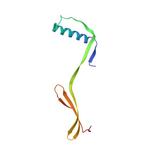
    5XFU

  • PubMed Abstract: 

    Rational engineering of a protein to enable domain swapping requires an understanding of the sequence, structural and energetic factors that favor the domain-swapped oligomer over the monomer. While it is known that the deletion of loops between β-strands can promote domain swapping, the spliced sequence at the position of the loop deletion is thought to have a minimal role to play in such domain swapping. Here, two loop-deletion mutants of the non-domain-swapping protein monellin, frame-shifted by a single residue, were designed. Although the spliced sequence in the two mutants differed by only one residue at the site of the deletion, only one of them (YEIKG) promoted domain swapping. The mutant containing the spliced sequence YENKG was entirely monomeric. This new understanding that the domain swapping propensity after loop deletion may depend critically on the chemical composition of the shortened loop will facilitate the rational design of domain swapping.


  • Organizational Affiliation
    • National Centre for Biological Sciences, Tata Institute of Fundamental Research, Bengaluru, 560065, India.

Macromolecule Content 

  • Total Structure Weight: 54.85 kDa 
  • Atom Count: 3,694 
  • Modeled Residue Count: 449 
  • Deposited Residue Count: 470 
  • Unique protein chains: 1

Macromolecules

Find similar proteins by:|  3D Structure
Entity ID: 1
MoleculeChains  Sequence LengthOrganismDetailsImage
Monellin chain B,Monellin chain A
A, B, C, D, E
94Dioscoreophyllum cumminsiiMutation(s): 0 
UniProt
Find proteins for P02881 (Dioscoreophyllum cumminsii)
Explore P02881 
Go to UniProtKB:  P02881
Find proteins for P02882 (Dioscoreophyllum cumminsii)
Explore P02882 
Go to UniProtKB:  P02882
Entity Groups
Sequence Clusters30% Identity50% Identity70% Identity90% Identity95% Identity100% Identity
UniProt GroupsP02881P02882
Sequence Annotations
Expand
Reference Sequence

Experimental Data & Validation

Experimental Data

  • Method: X-RAY DIFFRACTION
  • Resolution: 2.61 Å
  • R-Value Free:  0.294 (Depositor), 0.302 (DCC) 
  • R-Value Work:  0.260 (Depositor), 0.263 (DCC) 
  • R-Value Observed: 0.261 (Depositor) 
Space Group: C 1 2 1
Unit Cell:
Length ( Å )Angle ( ˚ )
a = 108.743α = 90
b = 87.265β = 127.85
c = 87.375γ = 90
Software Package:
Software NamePurpose
PHENIXrefinement
XDSdata reduction
XDSdata scaling
MOLREPphasing

Structure Validation

View Full Validation Report


Entry History 

& Funding Information

Deposition Data


Funding OrganizationLocationGrant Number
TIFR-DAEIndia--

Revision History  (Full details and data files)

  • Version 1.0: 2017-07-26
    Type: Initial release
  • Version 1.1: 2017-10-04
    Changes: Database references
  • Version 1.2: 2023-11-22
    Changes: Data collection, Database references, Refinement description