5X8N | pdb_00005x8n

Crystal structure of mouse importin-alpha1 bound to the nuclear localization signal of Epstein-Barr virus EBNA-LP protein


Experimental Data Snapshot

  • Method: X-RAY DIFFRACTION
  • Resolution: 2.15 Å
  • R-Value Free: 
    0.210 (Depositor), 0.211 (DCC) 
  • R-Value Work: 
    0.185 (Depositor), 0.186 (DCC) 
  • R-Value Observed: 
    0.186 (Depositor) 

Starting Model: experimental
View more details

wwPDB Validation 3D Report Full Report

Validation slider image for 5X8N

This is version 1.2 of the entry. See complete history

Literature

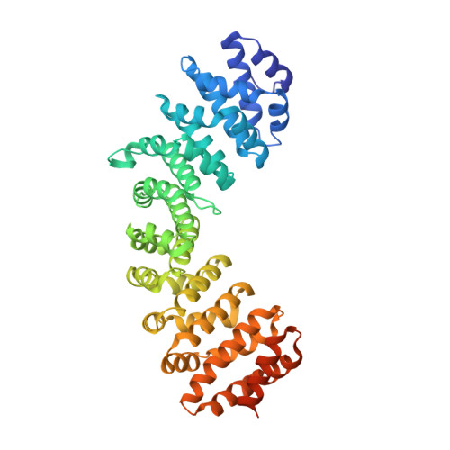
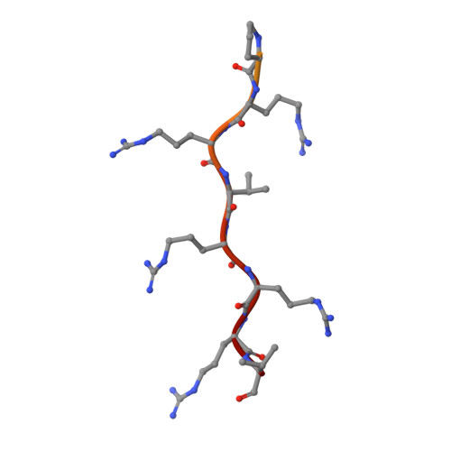
Crystal structure of importin-alpha bound to the nuclear localization signal of Epstein-Barr virus EBNA-LP protein

Nakada, R.Matsuura, Y.

(2017) Protein Sci 26: 1231-1235

  • DOI: https://doi.org/10.1002/pro.3173
  • Primary Citation Related Structures: 
    5X8N

  • PubMed Abstract: 

    Epstein-Barr virus EBNA-LP protein is a transcriptional coactivator of EBNA2. Efficient nuclear localization of EBNA-LP is essential for cooperation with EBNA2. Here, we report the crystal structure of the nuclear import adaptor importin-α1 bound to the nuclear localization signal (NLS) of EBNA-LP that shows EBNA-LP residues 44-RRVRRR-49 binding to the major NLS-binding site at the P0-P5 positions. In contrast to previously characterized classical NLSs that invariably have a basic residue [either lysine (in the vast majority of cases) or arginine] at the P2 position, the EBNA-LP NLS is unique in that it has valine at the P2 position. The loss of the critical P2 lysine (or arginine) is compensated by arginine at the P0 position in the EBNA-LP NLS.


  • Organizational Affiliation
    • Division of Biological Science, Graduate School of Science, Nagoya University, Japan.

Macromolecule Content 

  • Total Structure Weight: 52.59 kDa 
  • Atom Count: 3,439 
  • Modeled Residue Count: 433 
  • Deposited Residue Count: 482 
  • Unique protein chains: 2

Macromolecules

Find similar proteins by:|  3D Structure
Entity ID: 1
MoleculeChains  Sequence LengthOrganismDetailsImage
Importin subunit alpha-1460Mus musculusMutation(s): 0 
Gene Names: Kpna2Rch1
UniProt & NIH Common Fund Data Resources
Find proteins for P52293 (Mus musculus)
Explore P52293 
Go to UniProtKB:  P52293
IMPC:  MGI:103561
Entity Groups
Sequence Clusters30% Identity50% Identity70% Identity90% Identity95% Identity100% Identity
UniProt GroupP52293
Sequence Annotations
Expand
Reference Sequence
Find similar proteins by:  Sequence   |   3D Structure  
Entity ID: 2
MoleculeChains  Sequence LengthOrganismDetailsImage
Epstein-Barr nuclear antigen leader protein22Human herpesvirus 4 strain B95-8Mutation(s): 0 
UniProt
Find proteins for Q8AZK7 (Epstein-Barr virus (strain B95-8))
Explore Q8AZK7 
Go to UniProtKB:  Q8AZK7
Entity Groups
Sequence Clusters30% Identity50% Identity70% Identity90% Identity95% Identity100% Identity
UniProt GroupQ8AZK7
Sequence Annotations
Expand
Reference Sequence

Experimental Data & Validation

Experimental Data

  • Method: X-RAY DIFFRACTION
  • Resolution: 2.15 Å
  • R-Value Free:  0.210 (Depositor), 0.211 (DCC) 
  • R-Value Work:  0.185 (Depositor), 0.186 (DCC) 
  • R-Value Observed: 0.186 (Depositor) 
Space Group: P 21 21 21
Unit Cell:
Length ( Å )Angle ( ˚ )
a = 78.27α = 90
b = 90.29β = 90
c = 98.17γ = 90
Software Package:
Software NamePurpose
MOSFLMdata collection
Aimlessdata scaling
MOLREPphasing
PHENIXrefinement
PDB_EXTRACTdata extraction
Cootmodel building
Aimlessdata processing
MOSFLMdata reduction

Structure Validation

View Full Validation Report


Entry History 

Deposition Data

Revision History  (Full details and data files)

  • Version 1.0: 2017-04-19
    Type: Initial release
  • Version 1.1: 2017-06-07
    Changes: Database references
  • Version 1.2: 2023-11-22
    Changes: Data collection, Database references, Refinement description