5WRD

Crystal structure of LC3B in complex with FYCO1 LIR


Experimental Data Snapshot

  • Method: X-RAY DIFFRACTION
  • Resolution: 1.90 Å
  • R-Value Free: 0.244 
  • R-Value Work: 0.188 
  • R-Value Observed: 0.191 

wwPDB Validation   3D Report Full Report


This is version 1.1 of the entry. See complete history


Literature

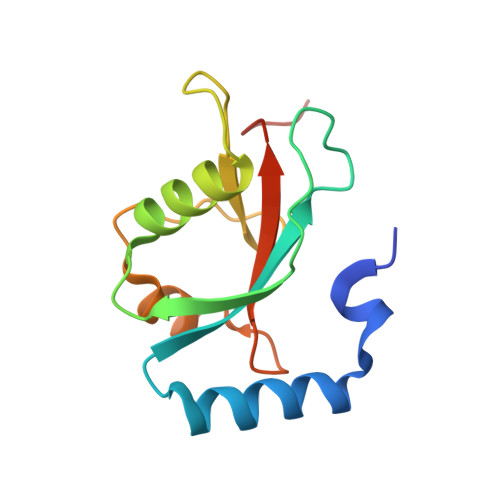
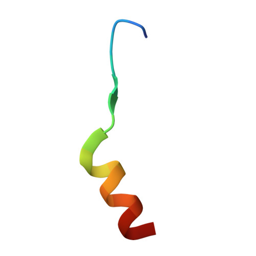
The crystal structure of mouse LC3B in complex with the FYCO1 LIR reveals the importance of the flanking region of the LIR motif

Sakurai, S.Tomita, T.Shimizu, T.Ohto, U.

(2017) Acta Crystallogr F Struct Biol Commun 73: 130-137

  • DOI: https://doi.org/10.1107/S2053230X17001911
  • Primary Citation of Related Structures:  
    5WRD

  • PubMed Abstract: 

    FYVE and coiled-coil domain-containing protein 1 (FYCO1), a multidomain autophagy adaptor protein, mediates microtubule plus-end-directed autophagosome transport by interacting with kinesin motor proteins and with the autophagosomal membrane components microtubule-associated protein 1 light chain 3 (LC3), Rab7 and phosphatidylinositol 3-phosphate (PI3P). To establish the structural basis for the recognition of FYCO1 by LC3, the crystal structure of mouse LC3B in complex with the FYCO1 LC3-interacting region (LIR) motif peptide was determined. Structural analysis showed that the flanking sequences N-terminal and C-terminal to the LIR core sequence of FYCO1, as well as the tetrapeptide core sequence, were specifically recognized by LC3B and contributed to the binding. Moreover, comparisons of related structures revealed a conserved mechanism of FYCO1 recognition by different LC3 isoforms among different species.


  • Organizational Affiliation

    Graduate School of Pharmaceutical Sciences, The University of Tokyo, 7-3-1 Hongo, Bunkyo-ku, Tokyo 113-0033, Japan.


Macromolecules
Find similar proteins by:  (by identity cutoff)  |  3D Structure
Entity ID: 1
MoleculeChains Sequence LengthOrganismDetailsImage
Microtubule-associated proteins 1A/1B light chain 3B
A, B
125Mus musculusMutation(s): 0 
Gene Names: Map1lc3bMap1alc3Map1lc3
UniProt
Find proteins for Q9CQV6 (Mus musculus)
Explore Q9CQV6 
Go to UniProtKB:  Q9CQV6
Entity Groups  
Sequence Clusters30% Identity50% Identity70% Identity90% Identity95% Identity100% Identity
UniProt GroupQ9CQV6
Sequence Annotations
Expand
  • Reference Sequence

Find similar proteins by:  Sequence   |   3D Structure  

Entity ID: 2
MoleculeChains Sequence LengthOrganismDetailsImage
Peptide from FYVE and coiled-coil domain-containing protein 1
C, D
19Mus musculusMutation(s): 0 
UniProt & NIH Common Fund Data Resources
Find proteins for Q8VDC1 (Mus musculus)
Explore Q8VDC1 
Go to UniProtKB:  Q8VDC1
IMPC:  MGI:107277
Entity Groups  
Sequence Clusters30% Identity50% Identity70% Identity90% Identity95% Identity100% Identity
UniProt GroupQ8VDC1
Sequence Annotations
Expand
  • Reference Sequence
Experimental Data & Validation

Experimental Data

  • Method: X-RAY DIFFRACTION
  • Resolution: 1.90 Å
  • R-Value Free: 0.244 
  • R-Value Work: 0.188 
  • R-Value Observed: 0.191 
  • Space Group: C 1 2 1
Unit Cell:
Length ( Å )Angle ( ˚ )
a = 113.412α = 90
b = 44.613β = 120.29
c = 63.582γ = 90
Software Package:
Software NamePurpose
REFMACrefinement
DENZOdata reduction
SCALEPACKdata scaling
MOLREPphasing

Structure Validation

View Full Validation Report


Entry History 

Deposition Data

Revision History  (Full details and data files)

  • Version 1.0: 2017-03-29
    Type: Initial release
  • Version 1.1: 2020-02-26
    Changes: Data collection