5WIA

Crystal structure of the segment, GNNSYS, from the low complexity domain of TDP-43, residues 370-375


Experimental Data Snapshot

  • Method: X-RAY DIFFRACTION
  • Resolution: 1.00 Å
  • R-Value Free: 0.181 
  • R-Value Work: 0.155 
  • R-Value Observed: 0.157 

wwPDB Validation   3D Report Full Report


This is version 1.6 of the entry. See complete history


Literature

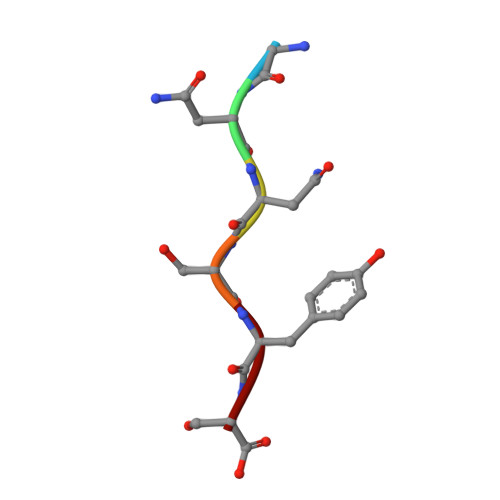
Atomic structures of TDP-43 LCD segments and insights into reversible or pathogenic aggregation.

Guenther, E.L.Cao, Q.Trinh, H.Lu, J.Sawaya, M.R.Cascio, D.Boyer, D.R.Rodriguez, J.A.Hughes, M.P.Eisenberg, D.S.

(2018) Nat Struct Mol Biol 25: 463-471

  • DOI: https://doi.org/10.1038/s41594-018-0064-2
  • Primary Citation of Related Structures:  
    5WHN, 5WHP, 5WIA, 5WIQ, 5WKB, 5WKD, 6CB9, 6CEW, 6CF4, 6CFH

  • PubMed Abstract: 

    The normally soluble TAR DNA-binding protein 43 (TDP-43) is found aggregated both in reversible stress granules and in irreversible pathogenic amyloid. In TDP-43, the low-complexity domain (LCD) is believed to be involved in both types of aggregation. To uncover the structural origins of these two modes of β-sheet-rich aggregation, we have determined ten structures of segments of the LCD of human TDP-43. Six of these segments form steric zippers characteristic of the spines of pathogenic amyloid fibrils; four others form LARKS, the labile amyloid-like interactions characteristic of protein hydrogels and proteins found in membraneless organelles, including stress granules. Supporting a hypothetical pathway from reversible to irreversible amyloid aggregation, we found that familial ALS variants of TDP-43 convert LARKS to irreversible aggregates. Our structures suggest how TDP-43 adopts both reversible and irreversible β-sheet aggregates and the role of mutation in the possible transition of reversible to irreversible pathogenic aggregation.


  • Organizational Affiliation

    Howard Hughes Medical Institute, University of California, Los Angeles, Los Angeles, CA, USA.


Macromolecules

Find similar proteins by:  Sequence   |   3D Structure  

Entity ID: 1
MoleculeChains Sequence LengthOrganismDetailsImage
TAR DNA-binding protein 436Homo sapiensMutation(s): 0 
UniProt & NIH Common Fund Data Resources
Find proteins for Q13148 (Homo sapiens)
Explore Q13148 
Go to UniProtKB:  Q13148
PHAROS:  Q13148
GTEx:  ENSG00000120948 
Entity Groups  
UniProt GroupQ13148
Sequence Annotations
Expand
  • Reference Sequence
Experimental Data & Validation

Experimental Data

  • Method: X-RAY DIFFRACTION
  • Resolution: 1.00 Å
  • R-Value Free: 0.181 
  • R-Value Work: 0.155 
  • R-Value Observed: 0.157 
  • Space Group: P 21 21 21
Unit Cell:
Length ( Å )Angle ( ˚ )
a = 4.786α = 90
b = 15.617β = 90
c = 40.679γ = 90
Software Package:
Software NamePurpose
DENZOdata reduction
SCALEPACKdata scaling
PHENIXrefinement
PDB_EXTRACTdata extraction
PHASERphasing

Structure Validation

View Full Validation Report


Entry History & Funding Information

Deposition Data


Funding OrganizationLocationGrant Number
National Institutes of Health/National Institute on Aging (NIH/NIA)United StatesNIH NIA AG029430

Revision History  (Full details and data files)

  • Version 1.0: 2018-04-25
    Type: Initial release
  • Version 1.1: 2018-05-30
    Changes: Data collection, Database references
  • Version 1.2: 2018-06-06
    Changes: Data collection, Database references
  • Version 1.3: 2018-06-20
    Changes: Data collection, Database references
  • Version 1.4: 2019-02-20
    Changes: Author supporting evidence, Data collection
  • Version 1.5: 2019-12-18
    Changes: Author supporting evidence
  • Version 1.6: 2024-03-13
    Changes: Data collection, Database references