5W3V

Crystal Structure of macaque APOBEC3H in complex with RNA


Experimental Data Snapshot

  • Method: X-RAY DIFFRACTION
  • Resolution: 2.24 Å
  • R-Value Free: 0.222 
  • R-Value Work: 0.182 
  • R-Value Observed: 0.184 

wwPDB Validation   3D Report Full Report


This is version 1.1 of the entry. See complete history


Literature

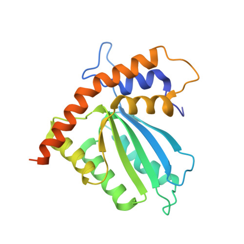
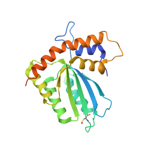
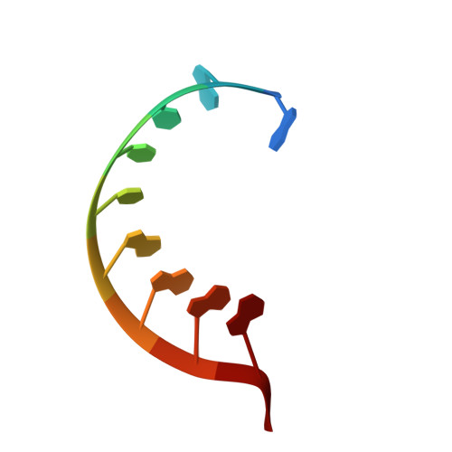
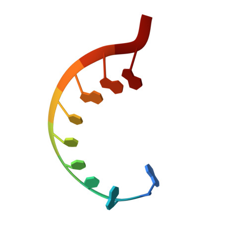
APOBEC3H structure reveals an unusual mechanism of interaction with duplex RNA.

Bohn, J.A.Thummar, K.York, A.Raymond, A.Brown, W.C.Bieniasz, P.D.Hatziioannou, T.Smith, J.L.

(2017) Nat Commun 8: 1021-1021

  • DOI: https://doi.org/10.1038/s41467-017-01309-6
  • Primary Citation of Related Structures:  
    5W3V

  • PubMed Abstract: 

    The APOBEC3 family of cytidine deaminases cause lethal hypermutation of retroviruses via deamination of newly reverse-transcribed viral DNA. Their ability to bind RNA is essential for virion infiltration and antiviral activity, yet the mechanisms of viral RNA recognition are unknown. By screening naturally occurring, polymorphic, non-human primate APOBEC3H variants for biological and crystallization properties, we obtained a 2.24-Å crystal structure of pig-tailed macaque APOBEC3H with bound RNA. Here, we report that APOBEC3H forms a dimer around a short RNA duplex and, despite the bound RNA, has potent cytidine deaminase activity. The structure reveals an unusual RNA-binding mode in which two APOBEC3H molecules at opposite ends of a seven-base-pair duplex interact extensively with both RNA strands, but form no protein-protein contacts. CLIP-seq analysis revealed that APOBEC3H preferentially binds to sequences in the viral genome predicted to contain duplexes, a property that may facilitate both virion incorporation and catalytic activity.


  • Organizational Affiliation

    Life Sciences Institute, University of Michigan, Ann Arbor, MI, 48109, USA.


Macromolecules

Find similar proteins by:  (by identity cutoff)  |  3D Structure
Entity ID: 1
MoleculeChains Sequence LengthOrganismDetailsImage
Apobec3H215Macaca nemestrinaMutation(s): 0 
UniProt
Find proteins for A0A1B2AH37 (Macaca leonina)
Explore A0A1B2AH37 
Go to UniProtKB:  A0A1B2AH37
Entity Groups  
Sequence Clusters30% Identity50% Identity70% Identity90% Identity95% Identity100% Identity
UniProt GroupA0A1B2AH37
Sequence Annotations
Expand
  • Reference Sequence
Find similar proteins by:  (by identity cutoff)  |  3D Structure
Entity ID: 2
MoleculeChains Sequence LengthOrganismDetailsImage
Apobec3H
B, C, D
215Macaca nemestrinaMutation(s): 0 
UniProt
Find proteins for A0A1B2AH37 (Macaca leonina)
Explore A0A1B2AH37 
Go to UniProtKB:  A0A1B2AH37
Entity Groups  
Sequence Clusters30% Identity50% Identity70% Identity90% Identity95% Identity100% Identity
UniProt GroupA0A1B2AH37
Sequence Annotations
Expand
  • Reference Sequence

Find similar nucleic acids by:  Sequence   |   3D Structure  

Entity ID: 3
MoleculeChains LengthOrganismImage
RNA (5'-R(P*AP*AP*CP*CP*CP*GP*GP*GP*GP*A)-3')
E, H
10synthetic construct
Sequence Annotations
Expand
  • Reference Sequence

Find similar nucleic acids by:  Sequence   |   3D Structure  

Entity ID: 4
MoleculeChains LengthOrganismImage
RNA (5'-R(P*AP*AP*CP*CP*CP*CP*GP*GP*GP*C)-3')
F, G
10synthetic construct
Sequence Annotations
Expand
  • Reference Sequence
Experimental Data & Validation

Experimental Data

  • Method: X-RAY DIFFRACTION
  • Resolution: 2.24 Å
  • R-Value Free: 0.222 
  • R-Value Work: 0.182 
  • R-Value Observed: 0.184 
  • Space Group: P 2 21 21
Unit Cell:
Length ( Å )Angle ( ˚ )
a = 87.96α = 90
b = 89.31β = 90
c = 134.37γ = 90
Software Package:
Software NamePurpose
PHENIXrefinement
Cootmodel building
XDSdata scaling
AutoSolphasing
XDSdata reduction

Structure Validation

View Full Validation Report


Entry History 

Deposition Data

Revision History  (Full details and data files)

  • Version 1.0: 2017-10-25
    Type: Initial release
  • Version 1.1: 2017-11-01
    Changes: Database references