Sulfur as an Acceptor to Bromine in Biomolecular Halogen Bonds.
Ford, M.C., Saxton, M., Ho, P.S.(2017) J Phys Chem Lett 8: 4246-4252
- PubMed: 28796521 Search on PubMed
- DOI: https://doi.org/10.1021/acs.jpclett.7b01725
- Primary Citation Related Structures:
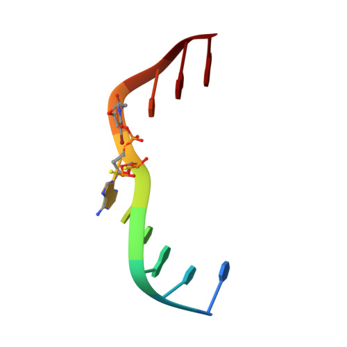
5VBJ - PubMed Abstract:
The halogen bond (X-bond) has become an important design element in chemistry, including medicinal chemistry and biomolecular engineering. Although oxygen is the most prevalent and best characterized X-bond acceptor in biomolecules, the interaction is seen with nitrogen, sulfur, and aromatic systems as well. In this study, we characterize the structure and thermodynamics of a Br···S X-bond between a 5-bromouracil base and a phosphorothioate in a model DNA junction. The single-crystal structure of the junction shows the geometry of the Br···S to be variable, while calorimetric studies show that the anionic S acceptor is comparable to or slightly more stable than the analogous O acceptor, with a -3.5 kcal/mol difference in ΔΔH 25 ° C and -0.4 kcal/mol ΔΔG 25 ° C (including an entropic penalty ΔΔS 25 ° C of -10 cal/(mol K)). Thus sulfur is shown to be a favorable acceptor for bromine X-bonds, extending the application of this interaction for the design of inhibitors and biological materials.
- Department of Biochemistry & Molecular Biology, Colorado State University , 1870 Campus Delivery, Fort Collins, Colorado 80523-1870, United States.
Organizational Affiliation: