5V7G

Crystal structure of NADPH-dependent glyoxylate/hydroxypyruvate reductase SMc04462 (SmGhrB) from Sinorhizobium meliloti in complex with NADPH and oxalate


Experimental Data Snapshot

  • Method: X-RAY DIFFRACTION
  • Resolution: 1.75 Å
  • R-Value Free: 0.180 
  • R-Value Work: 0.150 
  • R-Value Observed: 0.151 

wwPDB Validation   3D Report Full Report


Ligand Structure Quality Assessment 


This is version 1.4 of the entry. See complete history


Literature

Structural, Biochemical, and Evolutionary Characterizations of Glyoxylate/Hydroxypyruvate Reductases Show Their Division into Two Distinct Subfamilies.

Kutner, J.Shabalin, I.G.Matelska, D.Handing, K.B.Gasiorowska, O.Sroka, P.Gorna, M.W.Ginalski, K.Wozniak, K.Minor, W.

(2018) Biochemistry 57: 963-977

  • DOI: https://doi.org/10.1021/acs.biochem.7b01137
  • Primary Citation of Related Structures:  
    4WEQ, 4Z0P, 5J23, 5UNN, 5UOG, 5V6Q, 5V72, 5V7G, 5V7N

  • PubMed Abstract: 

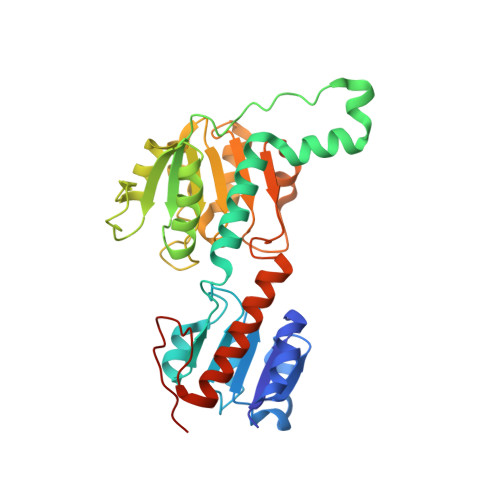
    The d-2-hydroxyacid dehydrogenase (2HADH) family illustrates a complex evolutionary history with multiple lateral gene transfers and gene duplications and losses. As a result, the exact functional annotation of individual members can be extrapolated to a very limited extent. Here, we revise the previous simplified view on the classification of the 2HADH family; specifically, we show that the previously delineated glyoxylate/hydroxypyruvate reductase (GHPR) subfamily consists of two evolutionary separated GHRA and GHRB subfamilies. We compare two representatives of these subfamilies from Sinorhizobium meliloti (SmGhrA and SmGhrB), employing a combination of biochemical, structural, and bioinformatics approaches. Our kinetic results show that both enzymes reduce several 2-ketocarboxylic acids with overlapping, but not equivalent, substrate preferences. SmGhrA and SmGhrB show highest activity with glyoxylate and hydroxypyruvate, respectively; in addition, only SmGhrB reduces 2-keto-d-gluconate, and only SmGhrA reduces pyruvate (with low efficiency). We present nine crystal structures of both enzymes in apo forms and in complexes with cofactors and substrates/substrate analogues. In particular, we determined a crystal structure of SmGhrB with 2-keto-d-gluconate, which is the biggest substrate cocrystallized with a 2HADH member. The structures reveal significant differences between SmGhrA and SmGhrB, both in the overall structure and within the substrate-binding pocket, offering insight into the molecular basis for the observed substrate preferences and subfamily differences. In addition, we provide an overview of all GHRA and GHRB structures complexed with a ligand in the active site.


  • Organizational Affiliation

    Department of Molecular Physiology and Biological Physics, University of Virginia , 1340 Jefferson Park Avenue, Charlottesville, Virginia 22908, United States.


Macromolecules
Find similar proteins by:  (by identity cutoff)  |  3D Structure
Entity ID: 1
MoleculeChains Sequence LengthOrganismDetailsImage
NADPH-dependent glyoxylate/hydroxypyruvate reductase
A, B, C, D
323Sinorhizobium meliloti 1021Mutation(s): 0 
Gene Names: SMc04462
EC: 1.1.1.81
UniProt
Find proteins for Q92LZ4 (Rhizobium meliloti (strain 1021))
Explore Q92LZ4 
Go to UniProtKB:  Q92LZ4
Entity Groups  
Sequence Clusters30% Identity50% Identity70% Identity90% Identity95% Identity100% Identity
UniProt GroupQ92LZ4
Sequence Annotations
Expand
  • Reference Sequence
Small Molecules
Ligands 5 Unique
IDChains Name / Formula / InChI Key2D Diagram3D Interactions
NDP
Query on NDP

Download Ideal Coordinates CCD File 
F [auth A],
K [auth B],
P [auth C],
T [auth D]
NADPH DIHYDRO-NICOTINAMIDE-ADENINE-DINUCLEOTIDE PHOSPHATE
C21 H30 N7 O17 P3
ACFIXJIJDZMPPO-NNYOXOHSSA-N
GOL
Query on GOL

Download Ideal Coordinates CCD File 
G [auth A],
L [auth B],
U [auth D]
GLYCEROL
C3 H8 O3
PEDCQBHIVMGVHV-UHFFFAOYSA-N
OXL
Query on OXL

Download Ideal Coordinates CCD File 
E [auth A],
J [auth B],
O [auth C],
S [auth D]
OXALATE ION
C2 O4
MUBZPKHOEPUJKR-UHFFFAOYSA-L
CL
Query on CL

Download Ideal Coordinates CCD File 
H [auth A],
M [auth B],
Q [auth C],
V [auth D]
CHLORIDE ION
Cl
VEXZGXHMUGYJMC-UHFFFAOYSA-M
NA
Query on NA

Download Ideal Coordinates CCD File 
I [auth A],
N [auth B],
R [auth C],
W [auth D]
SODIUM ION
Na
FKNQFGJONOIPTF-UHFFFAOYSA-N
Experimental Data & Validation

Experimental Data

  • Method: X-RAY DIFFRACTION
  • Resolution: 1.75 Å
  • R-Value Free: 0.180 
  • R-Value Work: 0.150 
  • R-Value Observed: 0.151 
  • Space Group: H 3
  • Diffraction Data: https://doi.org/10.18430/M35V7G
Unit Cell:
Length ( Å )Angle ( ˚ )
a = 178.209α = 90
b = 178.209β = 90
c = 133.799γ = 120
Software Package:
Software NamePurpose
MD2data collection
HKL-3000data reduction
HKL-3000data scaling
MOLREPphasing
REFMACrefinement
PDB_EXTRACTdata extraction
DENZOdata reduction
SCALEPACKdata scaling

Structure Validation

View Full Validation Report


Ligand Structure Quality Assessment 


Entry History & Funding Information

Deposition Data


Funding OrganizationLocationGrant Number
National Institutes of Health/National Institute of General Medical Sciences (NIH/NIGMS)United StatesGM094662
National Institutes of Health/National Institute of General Medical Sciences (NIH/NIGMS)United StatesGM117080
National Institutes of Health/National Human Genome Research Institute (NIH/NHGRI)United StatesHG008424

Revision History  (Full details and data files)

  • Version 1.0: 2017-03-29
    Type: Initial release
  • Version 1.1: 2017-09-13
    Changes: Author supporting evidence, Refinement description
  • Version 1.2: 2018-05-16
    Changes: Data collection, Database references, Structure summary
  • Version 1.3: 2019-11-27
    Changes: Author supporting evidence
  • Version 1.4: 2023-10-04
    Changes: Data collection, Database references, Derived calculations, Refinement description